The Problematic Genus Sclerocardius (Coleoptera: Curculionidae: Molytinae: Ithyporini)


Natural History Museum, Cromwell Road, London SW7 5BD, UK
Diversity 2018, 10(3), 74; https://doi.org/10.3390/d10030074
Submission received: 7 July 2018
/
Revised: 19 July 2018
/
Accepted: 20 July 2018
/
Published: 26 July 2018
(This article belongs to the Special Issue Systematics and Phylogeny of Weevils)
Abstract
:The genus Sclerocardius is revised, using morphological characters. Four species are recognized, including S. africanus (Boheman), S. bohemani Schoenherr stat.rev., S. indicus Hartmann and S. kuscheli sp.nov. The species Sclerocardius madecassus Ferragu is synonymized with S. bohemani syn.nov., and Charactocnemus hintzi Hartmann is treated as a junior synonym of S. bohemani, not S. africanus. A key to species is given. Lectotypes are designated for Heteramphus africanus Boheman and Sclerocardius africanus Schoenherr. A female elytro-tergal stridulatory system involving the modification of the wing-binding patch of the seventh tergite is reported for the Sclerocardiina for the first time and supports the inclusion of the subtribe within the Ithyporini.
1. Introduction
The genus Sclerocardius is an odd-looking weevil. With a huge rounded pronotum, elongate flanges on the fore tibiae, and a very narrow rostrum it looks like nothing else. Although it has been placed in the Ithyporini since 1935, this conveyed little about its relationships, since until relatively recently the tribe has been rather a dumping ground for taxa with a prosternal canal but no mesoventrite receptacle. The treatment by Alonso-Zarazaga & Lyal [1] as a subtribe of Ithyporini, followed by that of Lyal [2] as a separate tribe, indicates the continuing uncertainty as to its relationships. That is resolved in this paper.
There is some uncertainty about the names to be applied to the African species of Sclerocardius. The earlier-described species, Heteropus africanus Boheman, 1845, Sclerocardius bohemani Schoenherr, 1847 and Charactocnemus hintzi Hartmann, 1896, had all been synonymised and from 1897 have been known as S. africanus. Ferragu [3] described a new species, Sclerocardius madecassus from Madagascar, distinguishing it from S. africanus by external and internal characters. However, examination of a number of specimens from African countries identified as S. africanus revealed specimens with the same characters as described for S. madecassus, suggesting that this species is much more widely distributed than expected. This leads to a problem regarding which name to apply to which species. None of the descriptions of S. africanus, H. bohemani and C. hintzi provide sufficient information to distinguish between the two species which are hitherto known to occur, and the geographical distribution overlaps, so no name could be assigned with full confidence to any of the species. This problem is addressed below.
In addition to the two known African species a further species has been found in Angola. This is described below.
2. Materials and Methods
Descriptions are made on external and internal characters. Adult specimens were examined using a Zeiss SV11 stereomicroscope with a magnification of up to 6.4 and a ×2 additional objective; dissected genitalia were examined using this and a Laborlux 12 compound microscope. Drawings were made using a camera lucida on each microscope. To examine the genitalia, specimens were relaxed in de-ionised water and heated on a Tecam Dri-Block DB-1. The abdomen was removed and warmed in 10% KOH solution to macerate the internal tissues. Following maceration, the abdomen was transferred to de-ionised water and the genitalia dissected out of the abdomen. After resting in the water to wash off the KOH the genitalia were transferred to glycerol for imaging and preservation, and the abdomen glued flat on a card pinned beneath the specimen, with the terga and sterna both visible. The genitalia were transferred into glycerol for imaging and preservation. Following examination and imaging, the dissected genitalia are preserved in glass microvials pinned beneath the rest of the specimen. Habitus photographs were taken using a Canon 5 dsR camera with a 100 mm macro lens. Terminalia were placed in a cavity microscope slide with glycerol or KY gel, and photographs were taken using a Canon EOS 55D camera attached to a Leica 125 stereomicroscope. Habitus and terminalia images were stacked using Helicon Focus stacking software. Images were cleaned using GIMP 2.8 software.
Specimen label transcriptions are written verbatim; lines on a label are separated by a slash: “/” (type specimens only) and different labels by the word “and”.
Specimen length measurements were taken in lateral view along a straight line between the anterior and posterior of the structures concerned (see Figure 6 of Lyal & Curran [4]); the elytral length was taken from the anterior of the scutellum to the posterior extent of the elytra, the total length from the front of the head capsule to the posterior of the elytra. The pronotal width and elytral width refer to the maximum width of each. The morphological terminology follows Lyal [5].
The material examined is housed in the following collections, identified by the following codens:
- BMNH—Natural History Museum, London, United Kingdom
- NHRS—Naturhistoriska Riksmuseet, Stockholm, Sweden.
- MTD—Museum für Tierkunde, Dresden, Germany
3. Taxonomy
3.1. Genus Sclerocardius Schoenherr, 1847
Sclerocardius Schoenherr, 1847:82 [6]
Type species: Sclerocardius bohemani Schoenherr, 1847 (original designation, combined description). Gender: Masculine
= Heteropus Schoenherr, 1845: 1 [7] (non Palisot de Beauvois, 1820, nec Fitzinger, 1826, nec Jourdan, 1837, nec Spinola, 1837, nec Germar, 1839, nec Laporte, 1840, nec Hodgson, 1843). (synonymised with Sclerocardius by Lacordaire 1865: 317 [8]).
Type species: Heteropus africanus Boheman, 1845 (original designation)
Type species: Charactocnemus hintzi Hartmann, 1896 (monotypy) = Sclerocardius bohemani Schoenherr, 1847
= Charactonemus: Hustache, 1936: 18 [11] (Unavailable name: Lapsus)
Description
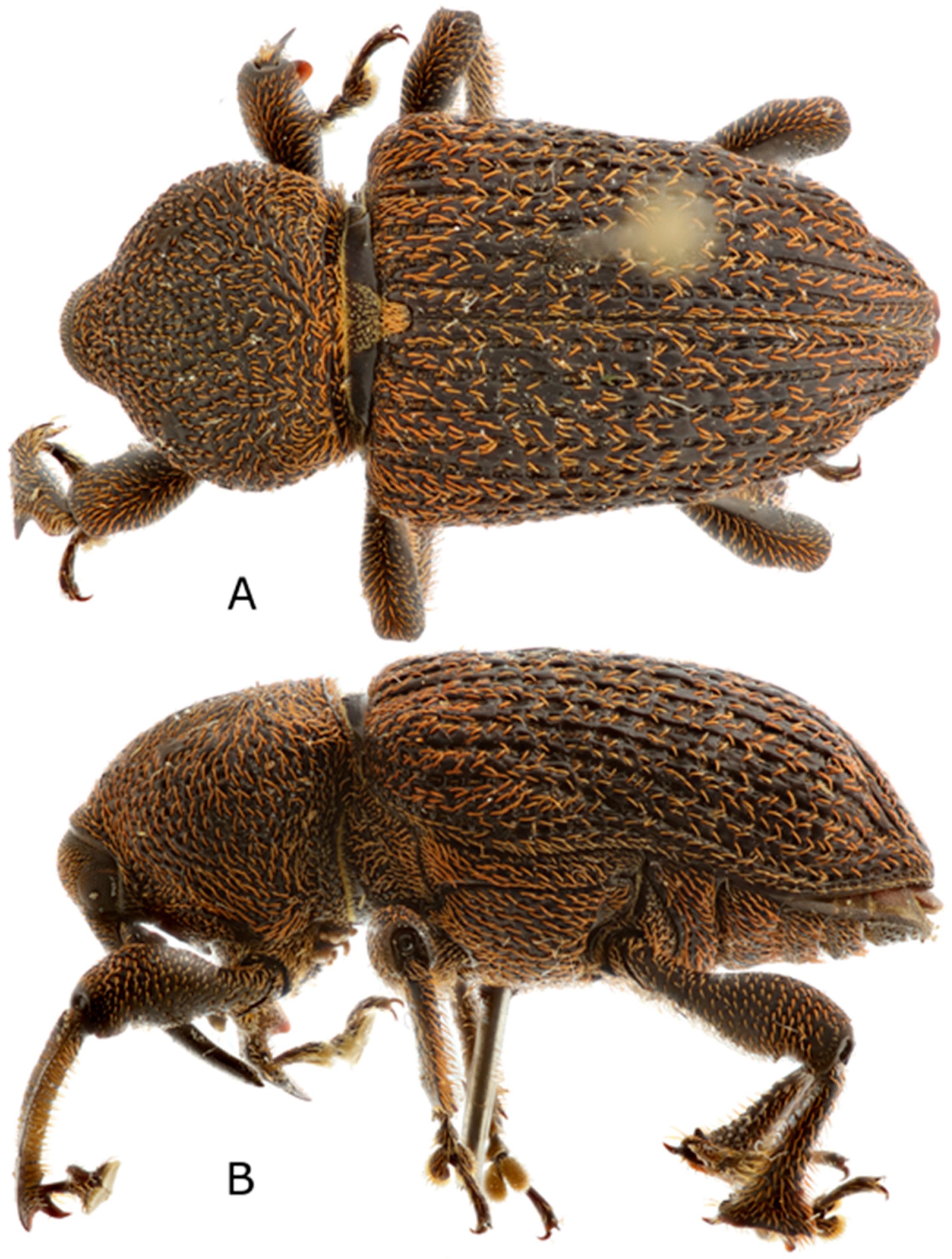
Large (6–14 mm long) weevils (Figure 1A,B, Figure 10A,B, Figure 14A,B and Figure 19A,B), with bulbous pronotum, fore tibiae with dorso-posterior flanges (Figure 6A, Figure 12A, Figure 15A and Figure 20A) and hind tibiae strongly expanded distally (Figures 6C, 12C, 15F and 20D).
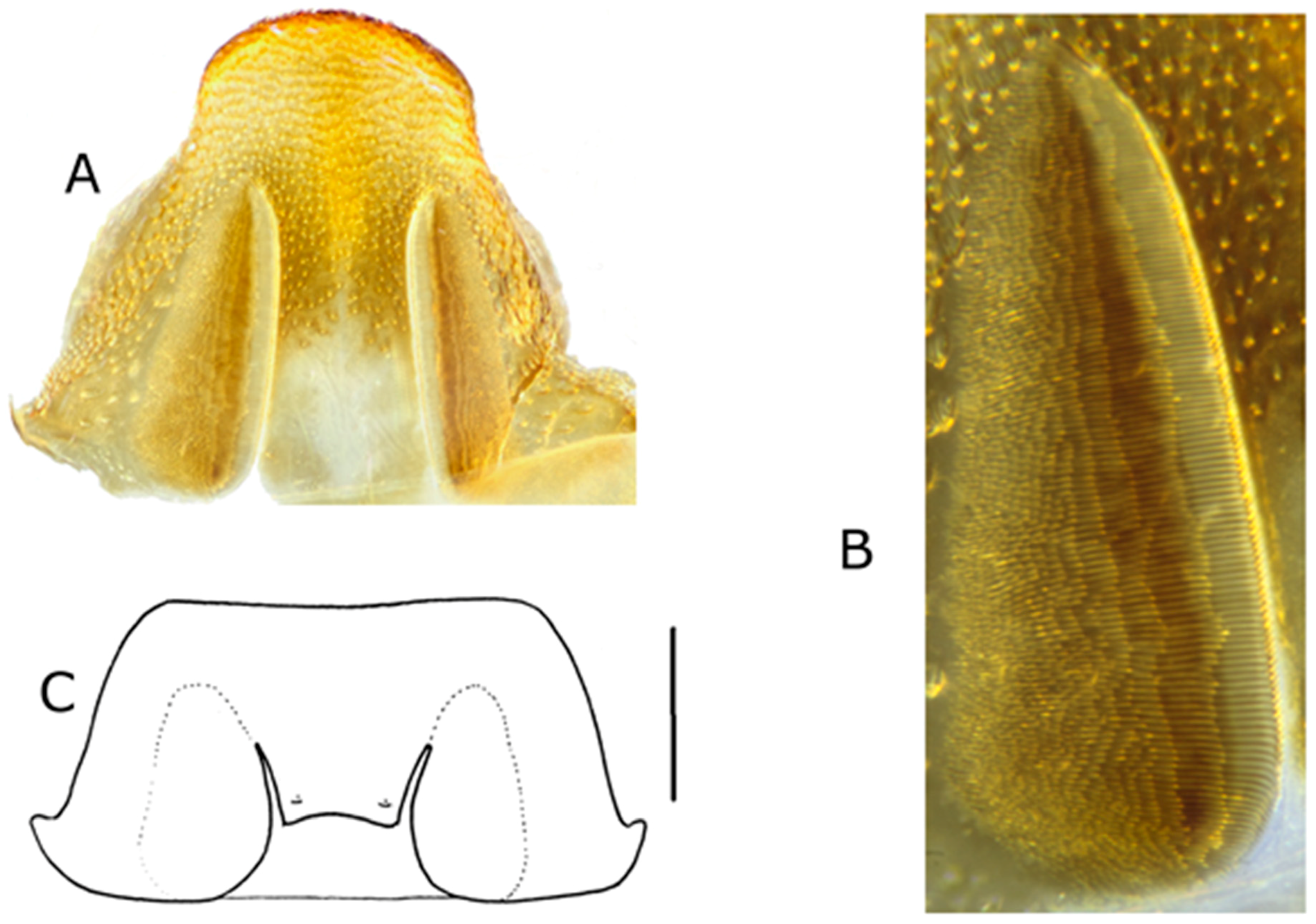
Head. Rostrum (Figure 2A,B) longer than prosternum, slender (approximate width of fore tibia omitting tibial teeth) and compressed. Mandibles lacking teeth on internal face, closing anteriorly to rostrum and projecting in a triangle (Figure 2A and Figure 3). Labial palps with three palpomeres. Scrobe not visible dorsally, slanting ventrally from anterior end about halfway along rostrum to underneath rostrum near eyes, scrobes separated by narrow carina under rostrum. Antennae (Figure 2C) with seven antennomeres in funicle, 3–5 quadrate, 6–7 transverse and broadening to club but not joining with club; club oval, slightly flattened, all club antennomeres with short dense pilosity, sutures sinuate. Eyes very large, lateral, extending slightly underneath rostrum but not approaching one another ventrally; ommatidia separately convex.
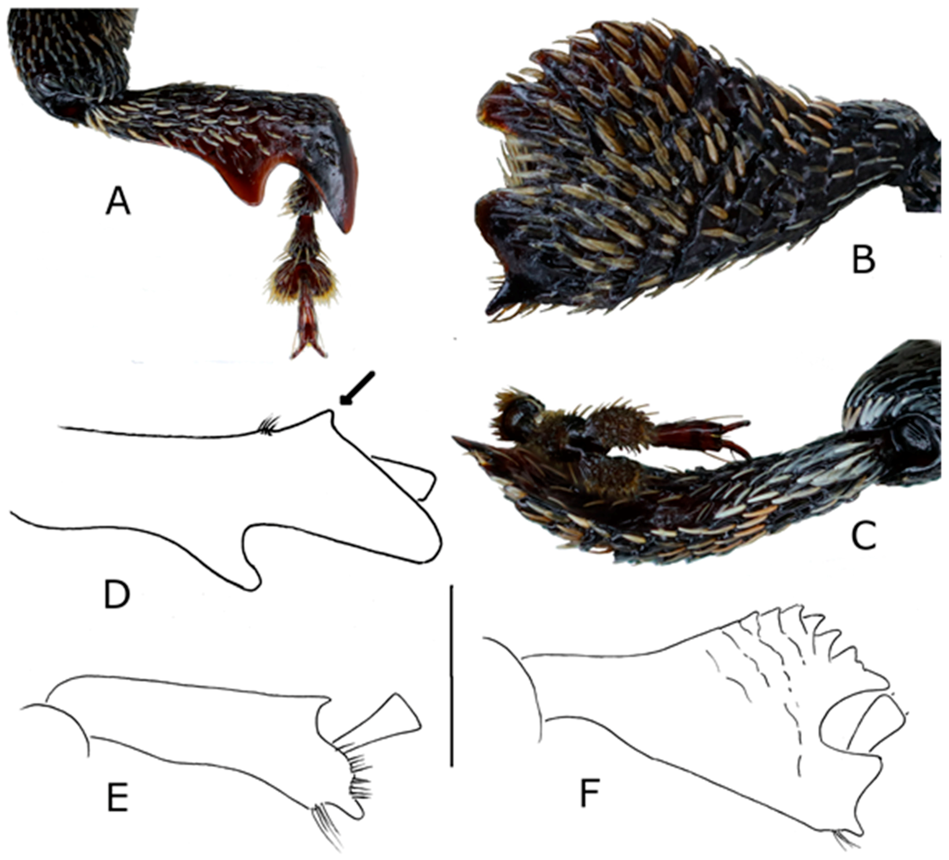
Thorax. Pronotum (Figures 1B, 10B, 14B and 19B) extending over head capsule, more or less convex dorsally in lateral aspect, more shallowly so in anterior aspect (Figure 3); notosternal suture well-marked, curved (Figure 2D). Prosternum (Figure 2D) with anterior margin emarginate; postocular lobes present; prothoracic rostral canal present, with lateral carina; procoxae separate, inner faces sometimes densely covered with long orange setae; sternellum depressed but post-coxal lamellae sometimes present and converging posteriorly immediately behind fore coxae to more or less close the gap between the coxae. Mesoventrite raised abruptly, anterior face weakly concave with weak lateral flanges directed anteriad. Metaventrite not depressed anteriorly; metanepisternal suture complete, sclerolepidia absent; metepimeron not fused to metanepisternum, concealed by elytron. Sclerolepidia absent. Elytra with basal margin concave; humeri developed; interstria III meeting interstria IX but not VI or VIII; submarginal fold pocket not broad; hind wings present, large. Elytro-tergal stridulatory structures present in male but not in females of all species; male elytral stridulatory file elongate and broad, near sutural margin. Femora with ventral tooth present, single, or absent. Fore tibia extended dorso-posteriorly into two large convex flattened lobes, one subapical and another medial, sometimes with a smaller prominence nearer the base (Figures 6A, 12A, 15A,D and 20A); uncus present, flattened; pre-mucro present; adventitious dorsal tooth sometimes present near apex; apical margin not distinct from inner flange, anterior setal comb absent; posterior apical setal comb on fore tibia curved round tarsal insertion. Mid tibia with premucro present or absent, uncus present, one or two dorsal adventitious teeth present apically, inner flange united with anterior apical margin and anterior apical setal comb on distal part of tibia. Hind tibiae with anterior apical setal comb broadened and developed into patch filling apical concavity between anterior apical margin and inner flange or, (S. kuscheli) not distinguishable from other setae and anterior apical margin not distinct from inner flange; inner flange where distinct developed into two or more flattened lobes (Figures 6C, 12C, 15F and 20D). Fifth tarsomere with ventral flat projection between and beneath base of claws. Tarsal claws simple, free and divaricate.
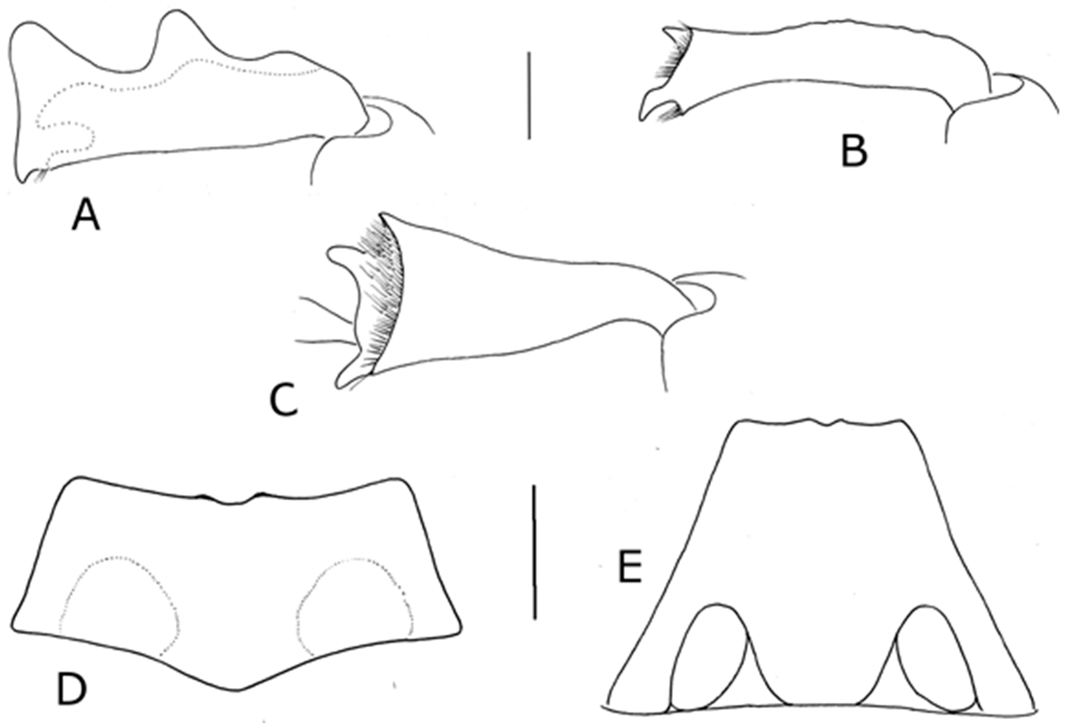
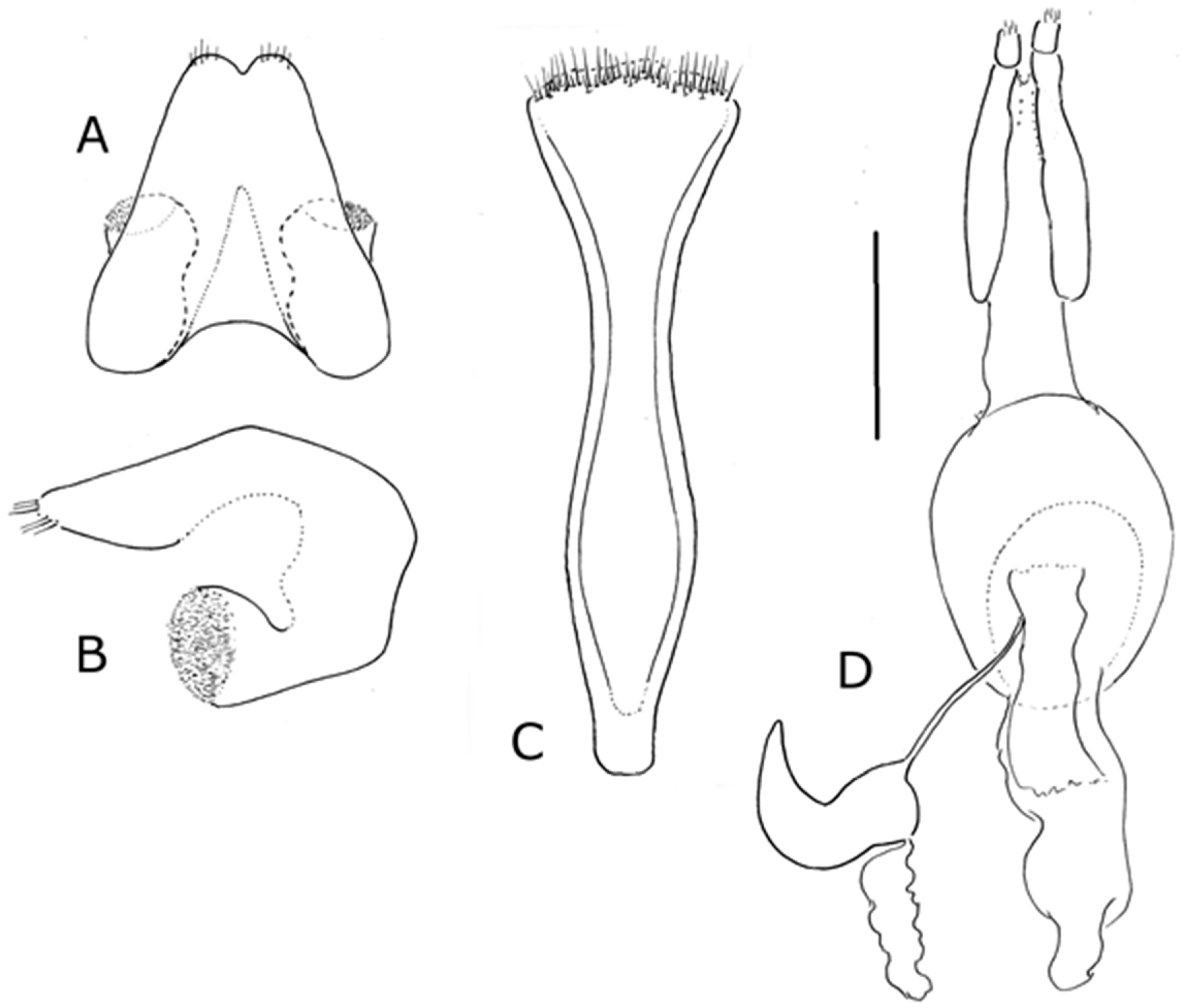
Abdomen. Abdominal tergites sclerotized. Pygidium not exposed in female; male apex of TVIII ventrally visible beyond emarginate posterior margin of ventrite 5. Male with elytro-tergal stridulation plectrum on tergite 7 as a pair of ridges on posterior margin (Figure 7A, Figure 12D and Figure 21A) or pair of plectral tubercles (Figure 16C); female wing-binding patches on tergite 7 not organised into file (Figures 7B, 12E and 21B) or with parallel arrangement on inner face (Figure 16A,B). Rectal loop present, weakly sclerotized, comprising six shallowly posteriorly-convex bands joining weak nodes.
Male terminalia. Sternite VIII entire, not separated into two hemisternites. Spiculum gastrale nearly straight, basal arms narrow and symmetrical. Tegmen with apodeme shorter or longer than diameter of ring; ring incomplete; parameroid lobes absent. Penis tubular, dorsally sclerotized, ostium opening posteriorly with apex extending posteriad ventrally (Figure 8D,E) or ostium opening more dorsally (Figure 17D,E), apodemes longer or shorter than body; endophallus lacking flagellum.
Female terminalia. Tergite VIII longer than wide, sides subparallel, posterior margin medially emarginate, convex on either side of emargination. Spiculum ventrale Y-shaped (Figure 9C and Figure 13H) or V-shaped, with basal arms subparallel, converging toward apex (Figure 18C and Figure 23C). Gonocoxites entire or with anterior unpigmented region, with elongate styli present; small tubular accessory gland sometimes present at base of each gonocoxite. Bursa at least as long as vagina, sometimes with large balloon-like anterior portion; membranous tubular extension from bursa to meet common oviduct (Figures 9E and 23D), or oviduct arising on membranous area separated from rest of bursa by sclerite (Figure 18D); spermathecal duct arising near common oviduct. Spermatheca with elongate conical sclerotized duct lobe, smaller conical sclerotized gland lobe.
Distribution
Angola, Democratic Republic of Congo, Ethiopia, Malawi, Mozambique, Nigeria, Senegal, Sierra Leone, South Africa, Swaziland, Tanzania, Togo, Zaire, Zambia, Zimbabwe; Madagascar; Sumatra.
Comments
Sclerocardius is the only genus placed in the subtribe Sclerocardiina. As discussed below, there is no other known genus that is morphologically similar, although there are indications that Ithyporus may be a close relative. The form of the body and the fore tibiae and the lack of a mesoventrite receptacle serve to distinguish species from any other currently known weevil.
Heteropus Schoenherr, 1845 is a junior homonym of Heteropus Palisot de Beauvois, 1805, leading Lacordaire 1865: 318 [8] to use the next available name, Sclerocardius Schoenherr, 1847. Schoenherr [7] attributed the genus Heteropus to Chevrolat, although there is no evidence that Chevrolat ever published the name. Subsequently, Schoenherr [6] stated that he had not seen Heteropus, so he may have taken the 1845 description from an unpublished source. However, authorship of the name rests with Schoenherr 1845.
In species where more extensive series are available for study, the uncus and sometimes premucro were much more acuminate in the younger specimens, suggesting wear with the age of the beetle. The form of the tibiae in particular suggests digging, and the large prothorax in particular suggests housing for enlarged leg muscles. The biology of adults and larvae is unknown, however.
Key to the species of Sclerocardius
- 1.
- Striae with large elongate foveae; interstriae lacking regular transverse creases (Figure 4A); hind tibia more than twice as long as maximum width, not strongly curved posteriad, anterior margin revealing internal flange (Figure 6C) ………………………………………………………………….2
- -
- Striae with no large foveae, interstriae with more or less regular transverse creases, giving an appearance of rectangular blocks (Figure 4B); hind tibia less than twice as long as maximum width, strongly curved posteriad, internal flange concealed by anterior margin (Figure 15B) Angola ………………………………………………………………….Sclerocardius kuscheli sp. nov.
- 2.
- Scales on pronotum absent, setiform or, if present and broader, less or only slightly longer than diameter of basal puncture and not or only just projecting above it (Figures 1A and 10A). Male with single small rounded tooth projecting from postero-ventral side of fore tibia (Figure 6A) (none in female). Africa, Madagascar. …………………………………………………………………3
- -
- Scales on pronotum and elytra bright orange, many at least twice as long as the diameter of the basal puncture and almost all projecting beyond it (Figure 19A). Male with several small rounded teeth projecting from postero-ventral side of fore tibia (Figure 20B) (none in female). Sumatra ………………………………………………………...……………. (Sclerocardius indicus Hartmann)
- 3.
- Pronotum punctate on disc, posteriorly punctures sometimes confluent and separated by raised irregular transverse ridges (Figure 1); penis body three-quarters the length of its apodemes, penis body length more than 2.6 times its maximum width (Figure 8D) Africa. ……………………………………………………………………. Sclerocardius africanus (Boheman)
- -
- Pronotum with raised irregular ridges between punctures posteriorly and extending onto disc (Figure 10A); penis body half the length of its apodemes, penis body length not more than 2.1 times its maximum width (Figure 13D) Africa, Madagascar. Sclerocardius bohemani Schoenherr
3.2. Sclerocardius africanus (Boheman, 1845)
Heteropus africanus Boheman in Schoenherr, 1845: 3 [7] (non H. africanus Palisot de Beauvois, 1805 [12])
Description
Figure 1, Figure 4A, Figure 5, Figure 6, Figure 7, Figure 8 and Figure 9.
Length 9.1–12.5 mm (mean 10.7 mm, n = 17); Pronotal width 2.9–5.5 mm (mean 4.6 mm, n = 17); Elytral width 3.5–6.56 mm (mean 5.6 mm, n = 17); males and females not significantly different in size.
Derm black, not developed into prominences or tubercles. Scales small, inconspicuous, pale or orange, rarely longer than the punctures in which they arise, not forming clear patterns. Setae longer and slender laterally on elytra and metathorax, on coxae, and ventrally on tibiae and femora (Figures 5 and 6A).
Head. Rostrum weakly curved, similar in males and females, strongly punctate laterally along most of its length, strongly to sparsely punctate dorsally in basal half and sometimes distal to antenna, dorsal surface basal to antennae smooth between punctures or weakly raised into irregular longitudinal rounded ridges, especially dorso-laterally; each puncture with dark short setiform elongate scale, most visible laterally in dorsal view; abruptly narrowed lateroventrally before eyes to form weak notch. Head capsule densely punctate dorsally, each puncture with a very small setiform scale.
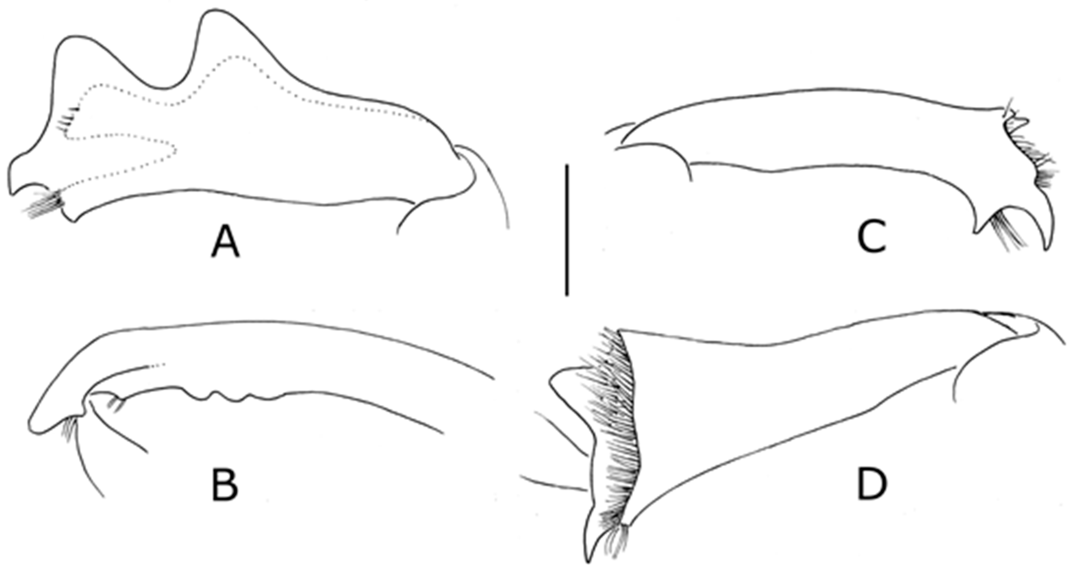
Thorax. Pronotum with length:width 0.83–1.00 (mean 0.89, n = 17), higher than long when length is taken as axis normal to height, strongly convex dorsally in lateral aspect (Figure 1B), more shallowly so in anterior aspect, punctate on disc with punctures separate, more posteriorly and laterally punctures sometimes confluent, and separated by irregular ridges running transversely on dorsum and more longitudinally laterally; anteriorly extending over head capsule (Figure 1A,B). Prothorax ventrally with deep narrow rostral canal with lateral carinae before fore coxae; fore coxae separate, with strong tuft of elongate orange scales on inner face (Figure 5); post-coxal lamellae present and converging posteriorly immediately behind fore coxae to more or less close the gap between coxae with bilobed wall. Elytra with length:width 1.1–1.40 (mean 1.34, n = 17); interstriae sometimes broad, sometimes distorted by very large strial punctures, these more or less rectangular (Figures 1A and 4A). Fore femora with small hooked femoral tooth in distal half (Figure 6A), other femora with smaller hook-like ventral tooth (Figure 6C). Fore tibia with postero-ventral tooth in male (Figure 6A), this absent in female; premucro prominent, uncus ventral on apex, acuminate in newly-emerged specimens, more rounded in older specimens, continuous with distal dorso-posterior lobe on posterior margin, dorso-posteriorly with three laminate asetose lobes, the distal two much larger than the basal one, which may be indistinct (Figure 6A). Mid tibia with dorsal margin somewhat irregular, premucro strong, long acuminate uncus and two dorsal apical teeth (Figure 6B). Hind tibial apex with premucro weak, inner flange bearing ventral acuminate uncus and more dorsal rounded tooth (Figure 6C).
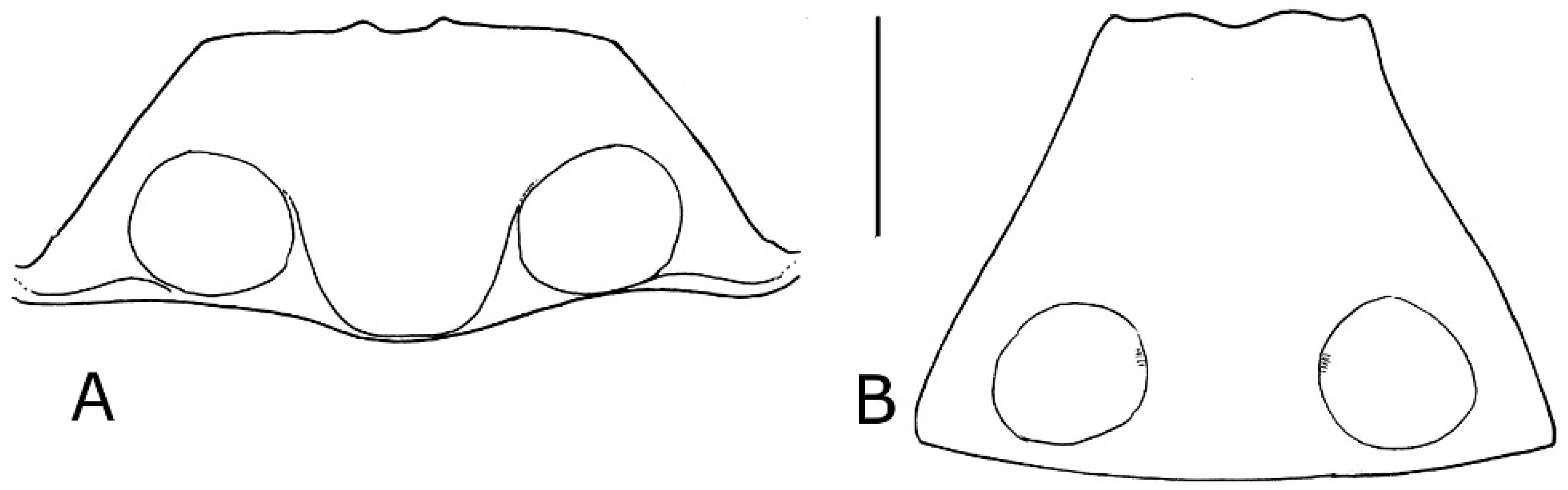
Abdomen. Tergites 1–6 weakly sclerotised; 7 more strongly sclerotised. Male tergite VII fairly evenly sclerotised; plectral tubercles with setae not present, but posterior margin with a pair of small raised ridges which may function as a plectrum (Figure 7A). Female tergite VII with posterior margin biconcave and median abrupt emargination (Figure 7B); spines of wing-binding patches not oriented in parallel along inner margin.
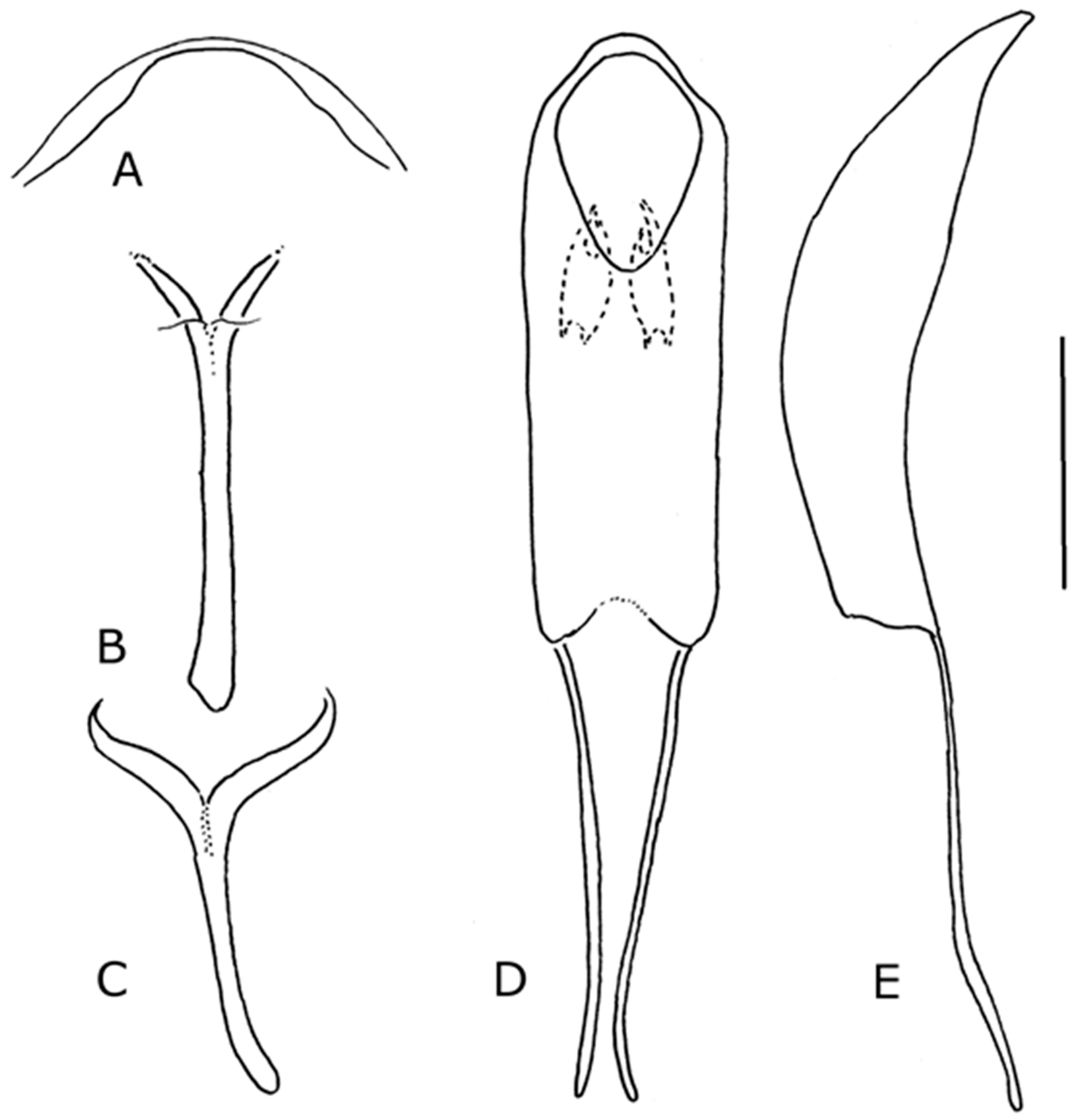
Male genitalia. Sternite VIII with two very weakly sclerotised lobes posteriorly, anterior of sclerite smoothly concave (Figure 8A). Spiculum gastrale Y-shaped, simple (Figure 8B). Tegmen with apodeme short, asymmetric (Figure 8C). Penis (Figure 8D–F) with body three-quarters the length of its apodemes, penis body length more than 2.6 times its maximum width; fully sclerotised; dorsally weakly concave; sides weakly convex anteriorly; ostium almost at right angles to long axis of penis body; small ventral projection anteriorly, anterior ventral margin with anteriad lobe, partially sclerotised (Figure 8F); endophallus with small cornet-shaped sclerite near gonopore
Female genitalia. Tergite VIII (Figure 9A) with posterior margin abruptly emarginate medially; approximately one-third as deep as wide (Figure 9B). Spiculum ventrale with posterior arms separate for two-thirds of length, with large membranous pouch between them, apodeme distinct anteriorly; posteriorly with numerous setae (Figure 9C). Gonocoxites entire, lacking median unpigmented area (Figure 9D). Vagina and bursa lacking pigmented area around junction with common oviduct and spermathecal duct; common oviduct and spermathecal duct arising separately from ventral membranous lobe of vagina (Figure 9E).
Distribution
South Africa, Sierra Leone, Ivory Coast, Togo, Zambia, Tanzania, Nigeria, Angola.
Comments
The characters distinguishing this species from S. bohemani are discussed under that species. Both species differ from S. kuscheli in having a much more convex pronotal profile, shorter dorsal scales, much broader elytral striae with large punctures, and a separate inner flange on the hind tibia. Sclerocardius indicus can be distinguished by the much longer orange scales.
The species was originally described under the homonymic genus name Heteropus. Although Heteropus africanus Boheman, 1845 has the same name string as Heteropus africanus Palisot de Beauvois, 1805, they are not homonyms, since they were not originally established in combination with the same generic name (Articles 53.3 and 57.8.1).
The earlier synonymies of Sclerocardius bohemani Schoenherr and Charactocnemus hintzi Hartmann are rejected here; this is discussed under S. bohemani below.
Boheman did not indicate the original number of specimens seen. Only one specimen has been located with the appropriate data to be in the type series, and it is designated as lectotype here.
Specimens examined
LECTOTYPE ♂, here designated, with the labels: “Senegallia/Chevrol.” [blue paper, handwritten] and [orange square] and “248.” [“148” printed but ‘1′ with handwritten ‘2′ superimposed, pink paper] and “♂“ [handwritten, white paper] and “7/83” [printed and handwritten, bright pink paper] and “Typus” [printed, red card] and “Riksmuseum / Stockholm” [printed, green paper] and “NHRS-GULI/000054255” [white card, printed] and “LECTOTYPE ♂ / Heteropus africanus / Boheman, 1845 / Lyal des. 2018” [printed, white paper] (NHRS). Other Material: South Africa: 1♂ with the labels “Natal” and “E. Gowring-Scopes Collection” and “BMNH(E) 2005-4”; 1♂ with the labels “Natal”and “23” and "Sclerocard. Bohemanni Sch. (sed Hete-ropi pertin et pot. and sp.)”; 1♀ with the labels “[indecipherable] Krantzkloof Natal” and “Distant Coll. 1911-383” and 1 ♂ with the labels “Pt. Natal” and “55.96”; 1♂ with the labels “Rustenburg, Transvaal” and “Rustenburg [indecipherable]” and “Distant Coll. 1911-383”. Angola: 1 ♂ with the labels “at light” and “Angola (A36) Chianga 21–24.iii.1972” and “Southern African Exp.” and “B.M. 1972-1” and “BMNH(E) 1237657”. Zambia: 1 ♂ with the labels “Zambia 1340m Jiwundu Swamp S11°51′54” E25°33′20” 21–24.ix.13 Light Trap. leg. Smith, R, & Takano, H. BMNH(E) 2013-71 1458902”. Tanzania: 1 ♂ with the label “Morogoro Tanganika territory 27.iii.21 N.C.E.Miller”. Sierra Leone: 1 ♂ with label “S. Leone Hill Station Nr. Freetown Oct. 1908 A. Pearse 1909-269”; 1 ♀ with the label “S. Leone 6771”; 2 ♂♂ with the labels “Sierra Leone Noala 17.viii.32 E. Hargreaves” and “G.A.K. Marshall Coll. B.M.1950-255”. Ivory Coast: 1 ♀ with labels “IVORY COAST 415m Telo Village outside Mt Sangbe NP 08°09′06.5”N 07°23′53.5”W” and “10-13.XI.15, Light Trap, Aristophanous, M., Moretto, P., Ruzzier, E. leg., BMNH(E) 2015-177” and “[QR code] 011218873”. Togo: 1 ♀ with the labels “Atakpalwé Togo West viii 1981 R.J. Cooter” and “Brit. Mus 1982-259”. Nigeria: 1 ♀ with the labels “M/V light White sheet” and “Nigeria Samaru 1–8. ix. 1970 P.H. Ward B.M. 1970-604”; 1 ♂ with the labels “M/V light White sheet” and “Nigeria Samaru 13–20. vii. 1970 P.H. Ward B.M. 1970-604”. West Africa: 1 ♂ Male: with the labels “W. Afr. Discove” and on the reverse “53 29”. Unknown: 1 ♂ with the label “Sclerocardius africanus Boh Nya[indicipherable]a”.
3.3. Sclerocardius bohemani Schoenherr, 1847
Sclerocardius bohemani Schoenherr, 1847: 84 [6]
Sclerocardius africanus; Lacordaire, 1865: 317 [8]
= Charactocnemus hintzi Hartmann, 1896: 185 [9], synonymised with S. africanus by (Hartmann 1897 [10]); here removed from that synonymy and synonymised with S. bohemani Schoenherr, 1847 syn. nov.
= Sclerocardius madecassus Ferragu, 1990: 107 [3] syn.nov.
Description
Figure 2, Figure 3, Figure 10, Figure 11, Figure 12 and Figure 13.
Length 8.64–14.16 mm (mean 11.69 mm, n = 32); Pronotal width 3.44–6.4 mm (mean 5.02 mm n = 32); Elytral width 4.48–7.68 mm (mean 6.18 mm, n = 32), males and females not significantly different in size.
Derm black, not developed into prominences or tubercles. Scales small, inconspicuous, pale or orange, rarely longer than the punctures in which they arise, not forming clear patterns. Setae longer and more slender laterally on elytra and metathorax, on coxae, and ventrally on tibiae and femora.
Head. Rostrum weakly curved, similar in males and females, strongly punctate laterally along most of length, strongly punctate dorsally in basal two-thirds, dorsal surface basal to antennae weakly raised into irregular longitudinal rounded carinae especially dorso-laterally, detail differing between specimens; each puncture with dark or pale short setiform scale; rostrum sometimes with weak notch before eyes. Head capsule densely punctate dorsally, each puncture with very small setiform scale.
Thorax. Pronotum with length:width 0.86–0.94 (mean 0.87, n = 32), higher than long when length is taken as axis normal to height (Figure 10B), strongly convex dorsally in lateral aspect (Figure 10B), more shallowly so in anterior aspect (Figure 3), punctate on disc with punctures sometimes confluent, and separated by irregular ridges running transversely on dorsum and more longitudinally laterally; anteriorly extending over head capsule. Prothorax ventrally with deep narrow rostral canal with lateral carinae before fore coxae; fore coxae separate, with strong tuft of elongate orange scales on inner face; post-coxal lamellae present and converging posteriorly immediately behind fore coxae to more or less close gap between coxae with bilobed wall. Elytra with length:width 1.22–1.40 (mean 1.32, n = 32); interstriae sometimes broad, sometimes distorted by very large strial punctures, these more or less rectangular. Fore femora with small hooked femoral tooth in distal half, other femora with very weak inconspicuous ventral tooth. Fore tibia (Figure 12A) with postero-ventral tooth in male (Figure 6A), this absent in female; premucro not prominent, uncus ventral on apex, acuminate, continuous with distal dorso-posterior lobe, three asetose dorso-posterior lobes, the distal two much larger than the basal one. Mid tibia (Figure 12B) with dorsal margin somewhat irregular, premucro strong, long acuminate uncus and two dorsal apical teeth, more anterior one weaker than in S. africanus. Hind tibial apex (Figure 12C) with premucro weak or undeveloped, inner flange bearing ventral acuminate uncus and more dorsal rounded tooth.
Abdomen. Male tergite VII (Figure 12D) fairly evenly sclerotised; plectral tubercles with setae not present, but posterior margin with pair of small raised ridges which may function as a plectrum. Female tergite VII (Figure 12E) with posterior margin medially and laterally emarginate; sometimes an indication of parallel rows of spines in anteromedial margin of wing-binding patch but these not obviously developed into plectrum.
Male genitalia. Sternite VIII with two very weakly sclerotised lobes posteriorly, anterior margin of sclerite tri-concave (Figure 13A). Spiculum gastrale Y-shaped, simple (Figure 13B). Tegmen with apodeme short, asymmetric (Figure 13C). Penis (Figure 13D,E) body half length of its apodemes, length not more than 2.1 times its maximum width; fully sclerotised; dorsally weakly concave; sides subparallel; ostium almost at right angles to long axis of penis body; small ventral projection anteriorly, anterior ventral margin with anteriad lobe, partially or completely sclerotised (Figure 13F,G); endophallus with pair of irregular small sclerites near gonopore.
Female genitalia. Very similar to those of Sclerocardius africanus and not figured separately. Tergite VIII with posterior margin abruptly emarginate medially; approximately one-third as deep as wide. Spiculum ventrale (Figure 13H) with posterior arms separate for two-thirds of length, with large membranous pouch between them, apodeme distinct anteriorly; posteriorly with numerous setae. Gonocoxites entire, lacking median unpigmented area. Vagina and bursa lacking pigmented area around junction with common oviduct and spermathecal duct; common oviduct and spermathecal duct arising separately from ventral membranous lobe of vagina.
Distribution
South Africa, Swaziland, Malawi, Angola, Zimbabwe, Ethiopia, Tanzania, Democratic Republic of Congo, Mozambique, N. Nigeria, Madagascar.
Comments
A male specimen from Tanzania differed from the other males dissected in having a slightly smaller penis (although the insect itself was of a similar size to the others) and the ventral anteriad projection of the penis body fully sclerotised and lanceolate (Figure 13F) rather than only sclerotised part-way and with an emarginate anterior margin (Figure 13E). The significance of this difference taxonomically is unclear and will require examination of additional specimens to be resolved.
Boheman [7] described Heteropus africanus from Senegal, stating that the type was in “Mus Dom Chevrolat”. Schoenherr [6] subsequently described Sclerocardius bohemani from “Montes Makkalisenses” in “Africa meridionali orientali” without having seen Heteropus africanus (although he was aware of the similarity). Lacordaire [8] subsequently synonymised S. bohemani Schoenherr with S. africanus (Boheman). Hartmann [9] described Charactocnemus hintzi, from “Ponguë bei Tanga, Deutsch-Ostafrika” (presumably the Tanga region, Tanzania) in 1896, but in the following year synonymised it with Sclerocardius africanus [10]. Most recently, Ferragu described Sclerocardius madecassus from Madagascar, differentiating it from S. africanus by the shorter penis and the dorsal sculpture, which is described as “tégument présentant sur les côtés et les bords de la face dorsale une forte rugosité produite par des rides profondes longitudinales, contiguës” (as opposed to “tégument lisse, brillant et pourvu de points petits et espacés”) [3].
Review of a number of specimens, and of the type material, has shown that the four names above represent two species, the most senior names for which are S. africanus and S. bohemani. Sclerocardius africanus has the pronotum punctate on the disc, the punctures posteriorly sometimes confluent and separated by raised irregular transverse ridges (Figure 1A), while the pronotum of S. bohemani has raised irregular ridges between the punctures both posteriorly and extending onto the disc (Figures 10A, 11); S. africanus has a penis body that is three-quarters the length of its apodemes and more than 2.6 times its maximum width (Figure 8D), while that of S. bohemani is half the length of its apodemes and not more than 2.1 times its maximum width (Figure 13D). The apical projections of the inner flange of the hind tibia in S. bohemani are less developed than those of S. africanus. The pronotal character is unequivocal in most specimens and has served to assign the types of all four species. The characters that separate S. bohemani from the other two species in the genus are the same as those already detailed for S. africanus.
Schoenherr did not indicate the original number of specimens seen when he described Sclerocardius bohemani. Only one specimen has been located with the appropriate data to be in the type series, and this is designated as lectotype here. The precise type locality cannot be identified. Wahlberg passed through the Magaliesberg on two trips: October 1841–August 1842 and June 1843 –December 1844 (Oberprieler, pers com), his routes being provided in his published journals [13].
Hartmann stated that he had only one specimen of Charactocnemus hintzi. Although not labeled with the original name, only one specimen has been found in the collection at Dresden that is of the correct genus and with the collection data quoted by Hartmann (Jäger, pers com).
Specimens examined
LECTOTYPE Sclerocardius bohemani Schoenherr 1847 ♀, here designated, with the labels: “Mont. Mak / kalisenses / Wahlberg.” [cream paper, handwritten] and [orange square] and “♀” [handwritten, white paper] and “140 / 83” [printed and handwritten, bright pink paper] and “Typus” [printed, red card] and “Riksmuseum / Stockholm” [printed, green paper] and, “NHRS-GULI / 000054254” [white card, printed] and “LECTOTYPE ♀ / Sclerocardius / bohemani / Schoenherr, 1847 / Lyal des. 2018” [printed, white paper] (NHRS).
HOLOTYPE Charactocnemus hintzi Hartmann 1896, with the labels: “Tauga / ostafrika / E. Hintz” [brown paper, handwritten] and “Sclerocardius / africanus Boh.” [brown paper, handwritten] and “Samm’ K.F. Hartmann / Ankauf 1941.1” [blue paper, printed] and “Staatl. Museum für / Tierkunde, Dresden” [white card, printed] and “HOLOTOTYPE / Charactocnemus / hintzi / Hartmann 1896 / Lyal vid. 2018” [printed, white paper] and “Sclerocardius / bohemani / Schoenherr 1847 / Lyal det. 2018” [printed, white paper] (MTD).
Other Material: South Africa: 1♀ with the labels “Port Natal” and on the reverse “49; 29”; 1♀ with the labels “Grahamstown” and “G.A.K. Marshall Coll. B.M. 1950-255”. Swaziland: 1♀ with the labels “Mt Chirunda Swaziland Swynnerton 1906” and “G.A.K. Marshall Coll. B.M. 1950-255” [this could be Mt Chirundu in Zimbabwe; a Mt Chirunda in Swaziland has not been located]. Zimbabwe: 1♀ with the label “Chirinda Rhodesia C.F.M.Swynnerton 1908-212”; 1♂ with the labels “Salisbury Dec 97 1884” and “Sharp Coll. B.M. 1948-336”; 1♀ with the label “Salisbury Mashonaland G.A.K. Marshall 1901-239”; 1♀ with the labels “Salisbury Mashonaland Jan 1901 G.A.K Marshall” and “G.A.K. Marshall Coll. B.M. 1950-255”; 1♀ with the labels “Salisbury Mashonaland Feb. 1899 G.A.K. Marshall” and “G.A.K. Marshall Coll. B.M. 1950-255”; 1♀ with the labels “Salisbury Mashonaland Dec. 1898 G.A.K. Marshall” and “G.A.K. Marshall Coll. B.M. 1950-255”; 1♀ with the label “Mashonaland G.A.K. Marshall 1908-212”; 1♀ with the label “N.W. Rhodesia Mwengwa 27°40′ E. 13° S 14.i.1914 H.C. Dollman” and on the reverse “on low shrubs about sundown”. Mozambique: 1♀ with the labels “Caia, Zambezi H. Swale 1913-117” and “23.2.12 Caia Zambezi H. Swale”; 1♀ with the labels “Delgoa H. Junod” and “G.A.K. Marshall Coll. B.M. 1950-255”; 1♀ with the label “Delagoa B”. Angola: 1♂ with the labels “Angola: Kwanza Norte prov., near N’Dalatando, collected at a petrol station 22.xi.2013, T. Lackner leg.” and “BMNH(E) 2014-40 T. Lackner”; 1♀ with the labels “Angola 19278” and “Angola Dundo 22.xii.1953 A.de B. Machado Pres by Com Inst Ent B.M. 1957-100”. Malawi: 2♀♀ 1♂ with the labels “Nyasaland 19 Karonga” and “E. Gowring-Scopes Collection BMNH(E) 2005-4”; 1♀ with the label “Nyasaland Mlanje 22.1.13 S.A. Neave 1914-123”. Democratic Republic of the Congo: 1♀ with the labels “Belgian Congo Mpala, Katanga 1-vii-1953 H. Bomans” and “E. Gowring-Scopes Collection BMNH(E) 2005-4” and “BMNH(E) # 716136 Digitally Imaged”; 1♀ with the labels “Quilu R. Congo” and “Sharp Coll. B.M. 1948-336”. Tanzania: 1♀ 1♂ with the labels “Tanganiyka: Old Shinyanga 1934 E. Burtt” and “Brit. Mus. 1935-257”; 1♂ with the labels “at light” and “Tanganyika Tanga Prov. iv–v. 1950 R.C.H.Sweeny B.M.1950-493”. Kenya: 1♀ with the labels “at light” and “Kenya Tanga, Ngomeni Mlingano Sisal Research Stn i-iii.1931” and “R.C.H Sweeny B.M. 1951-320”. Senegal: 1♀ with the labels “Seneg” and “Heteropus africanus Sch – Seneg” and “Bowring 63.47 *”. Nigeria: 2♀♀ with the labels “N. Nigeria Azare Dr. Ll. Lloyd” and “G.A.K. Marshall Coll. B.M. 1950-255”. Ethiopia: 1♀ with the label “Abyssinia Raffray” [probably high altitude, since this collector was collecting in the mountains]. Unknown locality: 1♂ with no labels; 1♀ with the label “G.A.K. Marshall Coll. B.M. 1950-255”; 1♀ with the labels “U[r]araga” and “D. Sharp Coll. B.M. 1932-116”.
3.4. Sclerocardius kuscheli sp. nov.
Description
Figures 4B and 14–18
Length 5.04–6.72 mm (mean 5.99 mm, n = 9); Pronotal width 2.0–2.88 mm (mean 2.54 mm, n = 9); Elytral width 2.4–3.28 mm (mean 2.91 mm, n = 9); males and females not significantly different in size.
Derm black, not developed into prominences or tubercles. Scales not concealing derm, elongate, white or yellowish- orange, longer than the punctures in which they arise, with white patch on either side of pronotum posteriorly, shading into orange anteriorly, elytral declivity with orange scales, more basally elytral scales pale; femoral scales pale, tibial scales more yellowish-orange.
Head. Rostrum weakly curved, similar in males and females; strongly punctate dorsally and laterally in basal half, each puncture with pale elongate scale, these longest dorsal to eyes; irregular longitudinal carinae sometimes present baso-laterally; abruptly narrowed before eyes lateroventrally to form weak indistinct notch. Head capsule densely punctate dorsally, each puncture with an elongate scale.
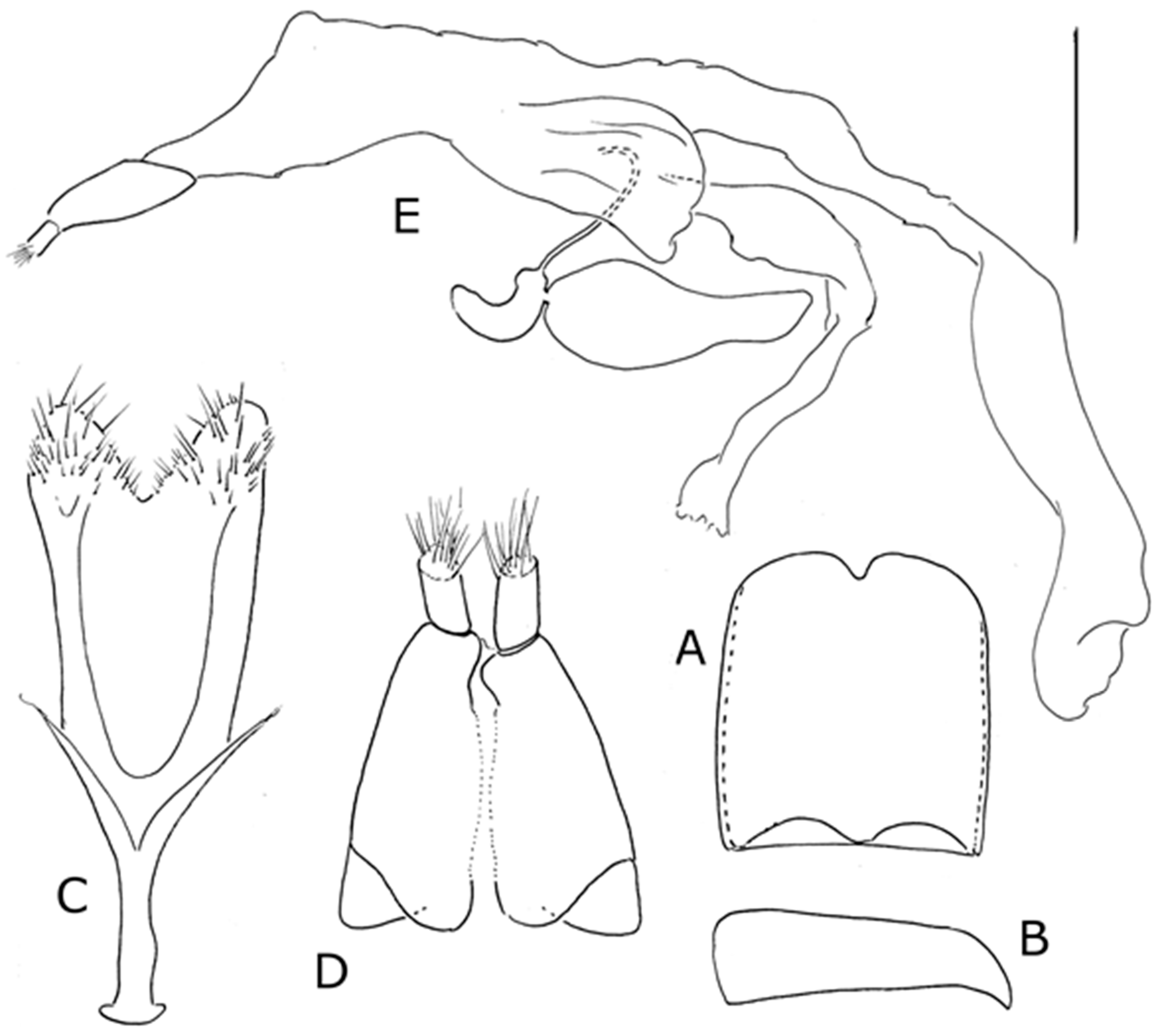
Thorax. Pronotum with length:width 0.70–0.77 (mean 0.75, n = 9), as high as long when length is taken as axis normal to height (Figure 14A), weakly convex dorsally in lateral aspect (Figure 14B), punctate on disc with punctures separate, without irregular ridges running transversely between them; anteriorly extending over head capsule. Prothorax ventrally with shallow narrow rostral canal with weak lateral carinae before fore coxae; fore coxae weakly separate, lacking tuft of elongate orange scales on inner face; post-coxal lamellae not developed. Elytra with length:width 1.39–1.50 (mean 1.45, n = 9); interstriae broad, with transverse rows of punctures, striae very narrow and linear, strial punctures narrow, more or less confluent (Figures 4B and 14A). Femoral teeth absent. Fore tibia lacking postero-ventral tooth in male and female; premucro present, small, uncus very small and forming part of distal posterior lobe although curved ventrad; dorso-posteriorly with two lobes, the distal one larger than the basal one (Figure 15A,D). Mid tibia (Figure 15E) with premucro well developed, uncus ventral, single dorsal adventitious tooth present; anterior apical setal comb on apical margin just dorsal to uncus. Hind tibia (Figure 15B,C,F) strongly broadened distally, anterior face with transverse ridges and apically inclined posteriad so that anterior apical margin with setal comb not distinguishable and inner flange continuous with anterior margin of the tibia; premucro undeveloped, uncus acuminate, apex with two rounded or acuminate teeth; dorsal margin apically with large laminate teeth continuous with apical teeth.
Abdomen. Tergites I-V weakly sclerotised, VI and VII more strongly sclerotised. Male tergite VII with posterior margin weakly emarginate, sclerotised area projecting anteriad between wing-binding patches bearing a pair of plectral tubercles near anterior margin (Figure 16C). Female tergite VII lacking plectral tubercles but with inner third of wing-binding patches with spines parallel, transverse, forming a series of ridges (Figure 16A,B).
Male terminalia. Sternite VIII narrowly sclerotised along posterior margin (Figure 17A). Spiculum gastrale Y-shaped, simple (Figure 17B). Tegmen (Figure 17C) with apodeme longer than width of ‘ring’, slender. Penis (Figure 17D,E) body weakly sclerotised dorsally, lack anteriad ventral lobe; ostium diagonal with respect to longitudinal axis of penis; endophallus with pair of oval toothed sclerites not near gonopore.
Female genitalia. Tergite VIII with ventrolateral bulbous lobe on either side directed posteriad and covered with microtrichiae (Figure 18A); dorsal plate with sides narrowing to setose posterior margin, emarginate medially (Figure 18B). Spiculum ventrale (Figure 18C) with no separate apodeme, but basal arms separate for whole length, the space between them being an open pocket opening within the genital chamber; posterior margin truncate. Gonocoxites lacking unsclerotized area. Posterior end of bursa with oval bulbous sclerite, the oviduct and spermathecal duct inserted ventrally in its membranous center (Figure 18D).
Distribution
Angola
Etymology
The species is named after my friend and mentor Willy Kuschel, a name particularly apposite since the new species has enabled the resolution of one troubling systematic problem while simultaneously producing a new one.
Comments
The new species S. kuscheli is placed in the genus because of the following synapomorphies: fore tibia with dorso-posterior margin produced into two laminate lobes; mid tibia with uncus flattened and with additional laminate projections; hind tibia broadened distally, with laminate projections distad; pronotum rounded in dorsal view. It differs from other members of the genus in the following characters: fore coxae lacking tuft of orange scales on inner face; penis body with ostium oblique (and of a type seen in many other Curculionidae) compared to more or less at right angles to the longitudinal axis of the penis, sometimes terminal and across the full diameter of the penis; female tergite VIII with unique lateroventral lobes; female tergite VII with wing-binding patches modified as a stridulatory file. It shares the form of the female spiculum ventrale with S. indicus, which also shows a very small patch of parallel spines on the wing binding patch of SVII in the females.
Sclerocardius kuscheli can be distinguished from all other species so far known in the genus by the form of the hind tibia, which is less than twice as long as deep, as opposed to much more than twice as long as deep, and has no clearly differentiated anterior apical margin and inner flange. It differs from the Oriental species by having pale and dark scales dorsally as opposed to orange ones, and from the other African species in having much longer dorsal scales and a much less convex pronotum. The aligned scales of the female tergite VII differentiate it from all other known species, as does the presence of plectral tubercles on the male tergite VII.
As noted, the wing-binding patches of tergite VII in the female S. kuscheli are modified as a file in a very similar way to that of Ithyporina, there being a longitudinal patch of spines on the inner margin that are elongate and parallel, although the patch is not as produced dorsad as much as in most species of that group [14]. Such a structure has been seen nowhere else in the Curculionidae, and supports the placement of Sclerocardius in the Ithyporini as restricted by Lyal [2]. The female genitalia of S. kuscheli resemble those of Ithyporus setulosus Hustache: the spiculum ventrale lacks an apodeme but instead the basal arms are separate for almost the entire length, meeting anteriorly and containing between them an open pocket opening into the genital chamber, and the common oviduct and spermathecal duct arise separately from a membranous area in the centre of a bowl-shaped sclerite at the base of the bursa (this area is more expanded than in S. kuscheli, and of a slightly different shape). The form of the spiculum ventrale is also shared with S. indicus, although that species has the female bursa of a similar form to that found in S. africanus and S. bohemani.
Specimens examined
HOLOTYPE, ♂ with the labels: “Holo- / type” [printed, white disc with red border] and “ANGOLA central / Bié prov., Kuemba env. / 23.xi.2012/T. Lackner leg.” [printed] and “BMNH (E) / 2014-48 / T. Lackner” [printed] and “HOLOTYPE / Sclerocardius / kuscheli Lyal, 2018 / Lyal det. 2018” [printed]. PARATYPES: 8 ♀♀ with same data as the holotype, but with “Para- / type” [printed, white disk with yellow border] and “PARATYPE / Sclerocardius / kuscheli Lyal / Lyal det 2018” [printed].
3.5. Sclerocardius indicus Hartmann, 1903
Sclerocardius indicus Hartmann, 1903: 29 [15]
Description
Figure 19, Figure 20, Figure 21, Figure 22 and Figure 23.
Length 7.9–11.2 mm (mean 9.94 mm, n = 10); Pronotal width 2.88–3.92 mm (mean 3.69 mm, n = 10); Elytral width 3.52–5.04 mm (mean 4.50 mm, n = 10), males and females not significantly different in size.
Derm black, not developed into prominences or tubercles. Scales not concealing derm, elongate and narrow, orange, longer than the punctures in which they arise (Figure 19A,B).
Head. Rostrum weakly curved, similar in males and females; strongly punctate laterally in basal half above scrobe and distally at same level as scrobe, weakly to strongly punctate dorsally basal to antennal insertion, very weakly punctate more distally, sometimes with rounded longitudinal ridges dorsally basally, each puncture with orange elongate setiform scale, longer than diameter of puncture; abruptly narrowed before eyes lateroventrally to form a notch. Head capsule strongly punctate dorsally, each puncture with an orange elongate setiform scale, longer than diameter of puncture.
Thorax. Pronotum with length:width 0.91–1.0 (mean 0.94, n = 10), as high as long when length is taken as axis normal to height (Figure 19B), weakly convex dorsally in lateral aspect (Figure 19B), punctate on disc with punctures separate or confluent, irregular rounded ridges running more or less antero-transversely between them, these ridges most pronounced laterally and posteriorly; anteriorly pronotum extends over head capsule (Figure 19A). Prothorax ventrally with broad rostral canal with lateral carinae before fore coxae; fore coxae separate, with tuft of elongate orange scales on inner face; post-coxal lamellae present and forming two transverse lobes immediately behind fore coxae to more or less close the gap between the coxae. Elytra with length:width 1.30–1.50 (mean 1.45, n = 10); interstriae with irregular transverse rows of punctures or lacking such rows, striae narrow and linear, strial punctures more or less confluent (Figure 19A). Fore femora with small hooked femoral tooth in distal half, other femora with smaller hook-like ventral tooth. Fore tibia (Figure 20A,B) with three rounded postero-ventral teeth in male, these absent in female; premucro prominent, uncus in ventral half of apex, acuminate, curved posteriad (Figure 20B), continuous with distal dorso-posterior lobe, dorso-posteriorly with two lobes, the distal one larger than the basal one. Mid tibia (Figure 20C) with premucro very large, acuminate, uncus acuminate, directed ventrad, two dorsal apical teeth, the more posterior one slightly larger than the more anterior. Hind tibia (Figure 20D) strongly broadened distally; apex with premucro very weak, inner flange bearing acuminate uncus and more dorsal rounded tooth.
Abdomen. Tergites I to VI weakly sclerotised. Male tergite VII (Figure 21A) lacking plectral tubercles, but with pair of prominences on posterior margin. Female TVII (Figure 21B) with very small patch of parallel transverse spines on inner margin of wing-binding patch.
Male genitalia. Sternite VIII narrow with very weakly sclerotised lobe posteriorly providing convex posterior margin to sternite (Figure 22A). Spiculum gastrale Y-shaped, simple (Figure 22D). Tegmen with apodeme short, asymmetric (Figure 22B). Penis body weakly sclerotised dorsally; sides concave posteriorly, convex anteriorly; ostium almost at right angles to long axis of penis body (Figure 22E,F); small ventral projection anteriorly, anterior ventral margin with long sclerotised anteriad lobe (Figure 22C); endophallus lacking internal sclerite.
Female genitalia. Tergite VIII with posterior margin abruptly and deeply emarginate medially (Figure 23A); approximately 0.4 times as deep as wide (Figure 23B). Spiculum ventrale (Figure 23C) with no separate apodeme, but basal arms separate for whole length, the space between them being an open pocket; posterior margin bilobed. Gonocoxites (Figure 23E) with small separately pigmented area ventrally anteriorly separated from main pigmented area. Common oviduct and spermathecal duct arising separately off a long lobe of the vagina and both distant from the bursa; vagina and bursa lacking internal sclerites (Figure 23D).
Distribution
Malaysia: Peninsular Malaysia, Sarawak.
Comments
Sclerocardius indicus can be distinguished from the other known species by the covering of elongate and sometimes very narrow orange scales; in all other species, scales are pale or dark and sometimes very small. The multiple postero-ventral rounded teeth on the fore tibia in the male are unique in the genus.
The genitalia show a mix between the morphologies shown by S. kuscheli and the other African species. The female spiculum ventrale has the same deep V form as S. kuscheli (and Ithyporus setulosus). However, the female sternite VII and the ovipositor itself are of a similar form to S. africanus and S. bohemani. The small patch of transverse spines on the wing-binding patch of the female TVII may suggest the loss of this character.
Specimens examined
PENINSULAR MALYSIA: ♂ with the labels “Malay Penin. Selangor. Bukit Kutu at light, 3500 ft. March 16th 1931 H.M. Pendlebury” and “Ex Coll F.M.S. Museum” on the reverse, and “Ex FMS Museum BM 1955-354”; 1♂ with the labels “Malay Penin. Ex: Coll. Ag. Dept. Bukit Fraser June 1921” and “ex coll. FMS Museum” on the reverse, and “Pres. By Imp. Inst. Ent. B.M. 1927-355”; 1♂ with the labels “Malay Penin. Larat Hills at light 3700ft, 8th Feb 1932 H.M. Pendlebury” and “Ex. Coll. F.M.S. Museum” on the reverse, and “Pres. By Com. Inst. Ent. B.M. 1952-94”; 1♂ with the labels “Malaysia, Pahang Tanah Rata, 4700ft. 3.x.-13.xi.1980” and “A. Harman Coll. B.M. 1982-49.”; 1♂ with the labels “Malay Penin. Kedah Peak, 3300ft. 22nd March 1928” and “at light H.M. Pendlebury Coll. F.M.S. Museums” on the reverse, and “Ex FMS Museum BM 1955-354”; 1♂ with the labels “69775”, “Perak” and “Fry Coll 1905-100”; 1♀ with the labels “Malay Penin. Perak F.M.S. ex coll. Perak Mus. C. Warang 19.” and “Ex FMS Museum BM 1955-354”; 1♀ with the labels “Malay Penin. Selangor. Bukit Kutu at light, 3500ft. April 18th 1926 H.M. Pendlebury” and “Ex Coll F.M.S. Museum” on the reverse, and “Ex FMS Museum BM 1955-354”; 1♀ with the labels “Malaya Tanah Rata 19.5.1939. H.T. Pagden, Agric. Dept. at light” and “G.A.K. Marshall Coll. B.M. 1930-255” and “G.2734”. SARAWAK: 1♂ with the labels “Guning Tamabo [indecipherable] Baram River Sarawak 7.11.1920 J. C. Moulton” and “Raffles Museum Singapore” on the reverse, “430” and “Pres. By Imp. Inst. Ent. B.M. 1941-7”.
4. Discussion: The Systematic Position of Sclerocardius
The adult insect is highly unusual in shape, with its expanded fore tibiae, strongly convex pronotum and relatively small head, and this has probably caused confusion as to the correct placement of the genus. Lacordaire [8] originally gave it a family-group name and placed it in the Sipalides, and it remained in the Sipalini until Marshall [16] transferred the genus to Ithyporinae (without mentioning the family-group name Sclerocardiini). Alonso-Zarazaga & Lyal [1] formally placed the Sclerocardiina as a subtribe of Ithyporini (although failed to note its earlier transfer by Marshall). Lyal [2] failed to find synapomorphies with the Ithyporini and referred to it both as a separate tribe and as a subtribe of Ithyporini. The examination in this study has revealed putative synapomorphic characters shared with Ithyporini in whole or in part, supporting its placement within this tribe.
Characters supporting the placement of Sclerocardius within Ithyporini include: mandibles smoothly convex exteriorly and narrow in lateral aspect, closing medially; antennal club with sutures sinuate; eyes extending under head (although not as far as in Ithyporini); prothoracic rostral canal bordered by strongly developed carinae; tarsal segment 5 extending beneath claws; tegmen unsclerotized dorsally and lacking parameroid lobes; female with wing-binding patch on TVII sometimes modified so that spines on inner edge are parallel and elongate, probably functioning as a stridulatory file. This last character is present in S. kuscheli only within the Sclerocardiina but is unique to Ithyporini and suggests homology.
Most species of Sclerocardius lack the apomorphic female stridulatory system of the Ithyporina, the female lacking both the modification to the wing binding patch on tergite VII (although there is sometimes an indication of regularity in the spines near the inner margin of the patch) or any indication of a stridulatory file on the elytron and plectral tubercles on tergite VII. However, the presence of the modified wing-binding patch in S. kuscheli suggests that the condition in other species of Sclerocardius represents a loss state.
Males of most species of Sclerocardius lack plectral tubercles on tergite VII, although they do have an elongate stridulatory file on the elytra. Instead, they appear to have a pair of protuberances on the posterior margin of the tergite, which may act as a plectrum. The exception to this is S. kuscheli, which has a pair of plectral tubercles medially on the tergite. Ithyporus setulosus has five pairs of plectral tubercles arranged in weakly converging lines, the more posterior tubercle lying on the posterior margin of the tergite.
There are some indications of a close relationship with Ithyporus, at least with I. setulosus Hustache, 1924. This species has the apex of the hind tibia elongate and scoop-shaped, although not as developed as in Sclerocardius. Perhaps significantly, the form of the female genitalia, particularly the junction of vagina, bursa, common oviduct and spermathecal duct, and the form of the spiculum ventrale, are very similar to the morphology of S. kuscheli, as discussed above.
Further work is required to place Sclerocardius more firmly, but for now it is retained as a subtribe of Ithyporini. This potentially creates a paraphyletic Ithyporina, and the placement needs to be examined more closely.
Sclerocardius appears strongly adapted for digging (although no observations have been published to support this assumption): fore tibia broadened with laminar projections; hind tibia with broad apex; fore coxae with trochanteral articulation on posterior face; prothorax very large and convex (suggesting enlarged muscles supporting forelegs); prosternum with prothoracic rostral canal bordered by carinae; rostrum narrow (suggesting that the head can be pulled down and the rostrum concealed while the insect digs). Whether this is in soil or plant material is not known. Other Ithyporini are known to bore as larvae into bark and sometimes into wood, although some species have been observed developing in seeds [2].
Sclerocardius contains four described species, three of which are found in the Afrotropical Region, one also in Madagascar, and the third in the Malaysian Peninsula and Sumatra. There is no doubt that S. indicus is congeneric with the other species, and seems to share some apomorphies with the two larger African species (male with ventro-posterior tibial teeth, female wing-binding patch on tergite VII unmodified, male tergite VII lacking plectral tubercles, penis with ostium nearly perpendicular), although this will need to be confirmed by further analysis. No specimens of the genus have been found in India. Within Africa, the broad distribution of both S. africanus and S. bohemani is surprising; there are some indications of variation within the latter species but research on far more specimens is required to evaluate this.
Funding
This research received no external funding.
Acknowledgments
I am very grateful to colleagues in several institutions who contributed information and advice. Miguel Alonso-Zarazaga of the Museo Nacional de Ciencias Naturales, Madrid, provided very helpful advice on the provisions of the Code of Zoological Nomenclature. A specimen from Ivory Coast was made possible thanks to the support of the African Natural History Research Trust (Hereford, UK) and Richard Smith. Olaf Jäger of the Museum of Zoology (Museum für Tierkunde) Senckenberg Natural History Collections Dresden kindly provided information on the holotype of Charactocnemus hintzi Hartmann and his colleague Marc Srour took the photographs reproduced in this paper as Figures 3 and 11. Gunvi Lindberg of the Swedish Museum of Natural History very helpfully provided information and photographs of the types of Sclerocardius bohemani Schoenherr and Heteropus africanus Boheman, the photographs being reproduced in this paper as Figures 1 and 10. Rolf Oberprieler provided useful information about Wahlberg’s collecting route in the 1840s. I am very grateful to Keita Matsumoto for Figures 14B, 16A and 16B. I am particularly grateful to Willy Kuschel for kindling my interest in Molytinae.
Conflicts of Interest
The author declares no conflict of interest.
References
- Alonso-Zarazaga, M.A.; Lyal, C.H.C. A World Catalogue of Families and Genera of Curculionoidea (Insecta: Coleoptera) Excluding (Scolytidae and Platypodidae); Entomopraxis: Barcelona, Spain, 1999. [Google Scholar]
- Lyal, C.H.C. Molytinae Schoenherr, 1832. In Coleoptera, Beetles. Morphology and Systematics. Volume 3. In Handbook of Zoology. Arthropoda: Insecta; Leschen, R.A.B., Beutel, R.G., Eds.; De Greuter: Berlin, Germany; Boston, MA, USA, 2014; pp. 529–570. [Google Scholar]
- Ferragu, M.; Richard, R. Trois Nouveaux Curculionides de Madagascar et de l’Île de La Réunion [Coleoptera]. Rev. Fr. Entomol. 1990, 12, 105–110. [Google Scholar]
- Lyal, C.H.C.; Curran, L.M. Seed-Feeding Beetles of the Weevil Tribe Mecysolobini (Insecta: Coleoptera: Curculionidae) Developing in Seeds of Trees in the Dipterocarpaceae. J. Nat. Hist. 2000, 34, 1743–1847. [Google Scholar] [CrossRef]
- Lyal, C.H.C. Glossary of Weevil Characters. Available online: http://weevil.info/glossary-weevil-characters (accessed on 25 June 2018).
- Schoenherr, C.J. Mantissa Secunda Familiae Curculionidum Seu Descriptiones Novorum Quorundam Generum Curculionidum; Typis Norstedt et Filii: Holmiae, Sweden, 1847. [Google Scholar]
- Schoenherr, C.J. Genera et Species Curculionidum, Cum Synonymia Hujus Familiae. Species Novae Aut Hactenus Minus Cognitae, Descriptionibus a Dom. Leonardo Gyllenhal, C. H. Boheman, et Entomologis Aliis Illustratae; Roret: Paris, France, 1845; Volume 8. [Google Scholar]
- Lacordaire, T. Toire Naturelle Des Insectes. Genera Des Coléoptères Ou Exposé Méthodique et Critique de Tous Les Genres Proposés Jusqu’ici Dans Cet Ordre D’insectes; Librairie Encyclopédique de Roret: Paris, France, 1865; Volume 7. [Google Scholar]
- Hartmann, F. Beschreibung Einer Neuen Gattung Der Rüsselkäfer Aus Deutsch-Ostafrika. Dtsch. Entomol. Z. 1896, 1896, 185–188. [Google Scholar] [CrossRef]
- Hartmann, F. Neue Rüssel-Käfer Aus Deutsch-Ostafrika. Dtsch. Entomol. Z. 1897, 1, 81–96. [Google Scholar] [CrossRef]
- Hustache, A. Curculionidae: Cryptorrhynchinae. In Coleopterorum Catalogus Auspiciis et Auxilio W. Junk. 151; Schenkling, S., Ed.; Junk: Gravenage, The Netherlands, 1936; 317p. [Google Scholar]
- Palisot de Beauvois, A.M. Insectes Recueillis En Afrique et En Amerique, Dans Les Royaumes d’Oware et de Benin, à Saint-Dominique et Dans Les Etats-Unis, Pendant Les Années 1786–1797; Levrault: Paris, France, 1805. [Google Scholar]
- Wahlberg, J.; Craig, A.; Hummel, C. Johan August Wahlberg: Travel Journals (and Some Letters) South Africa and Namibia/Botswana, 1838–1856; Van Riebeck Society: Cape Town, South Africa, 1994. [Google Scholar]
- Lyal, C.H.C.; King, T. Elytro-Tergal Stridulation in Weevils (Insecta: Coleoptera: Curculionoidea). J. Nat. Hist. 1996, 30, 703–773. [Google Scholar] [CrossRef]
- Hartmann, F. Neue Exotische Rüsselkäfer. Wiener Entomol. Zeitung 1903, 22, 23–30. [Google Scholar]
- Marshall, G.A.K. New Curculionidae (Col.) from Tropical Africa, with Notes on Synonymy Etc. Ann. Mag. Nat. Hist. 1935, 10, 497–518. [Google Scholar] [CrossRef]
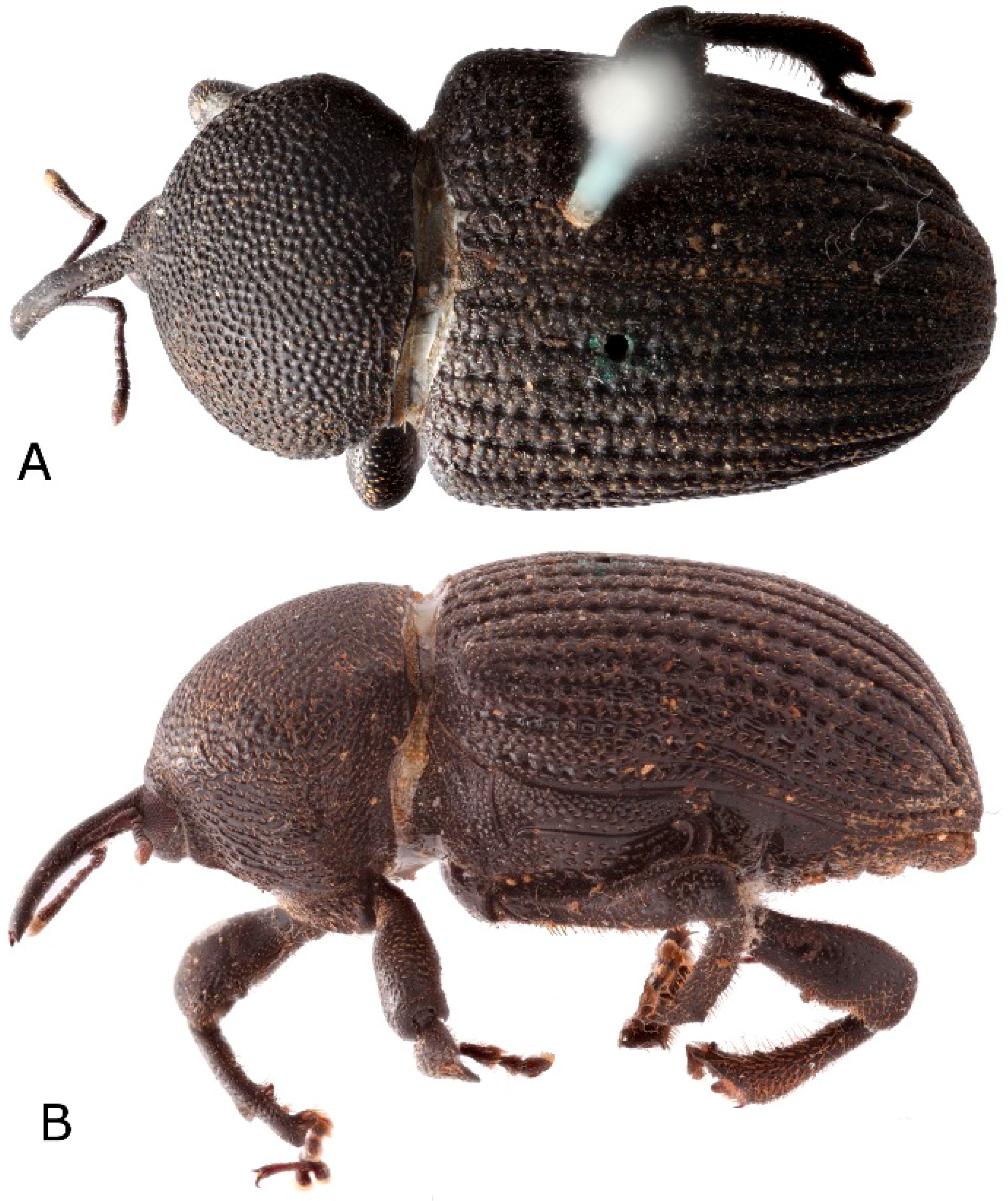
Figure 1.
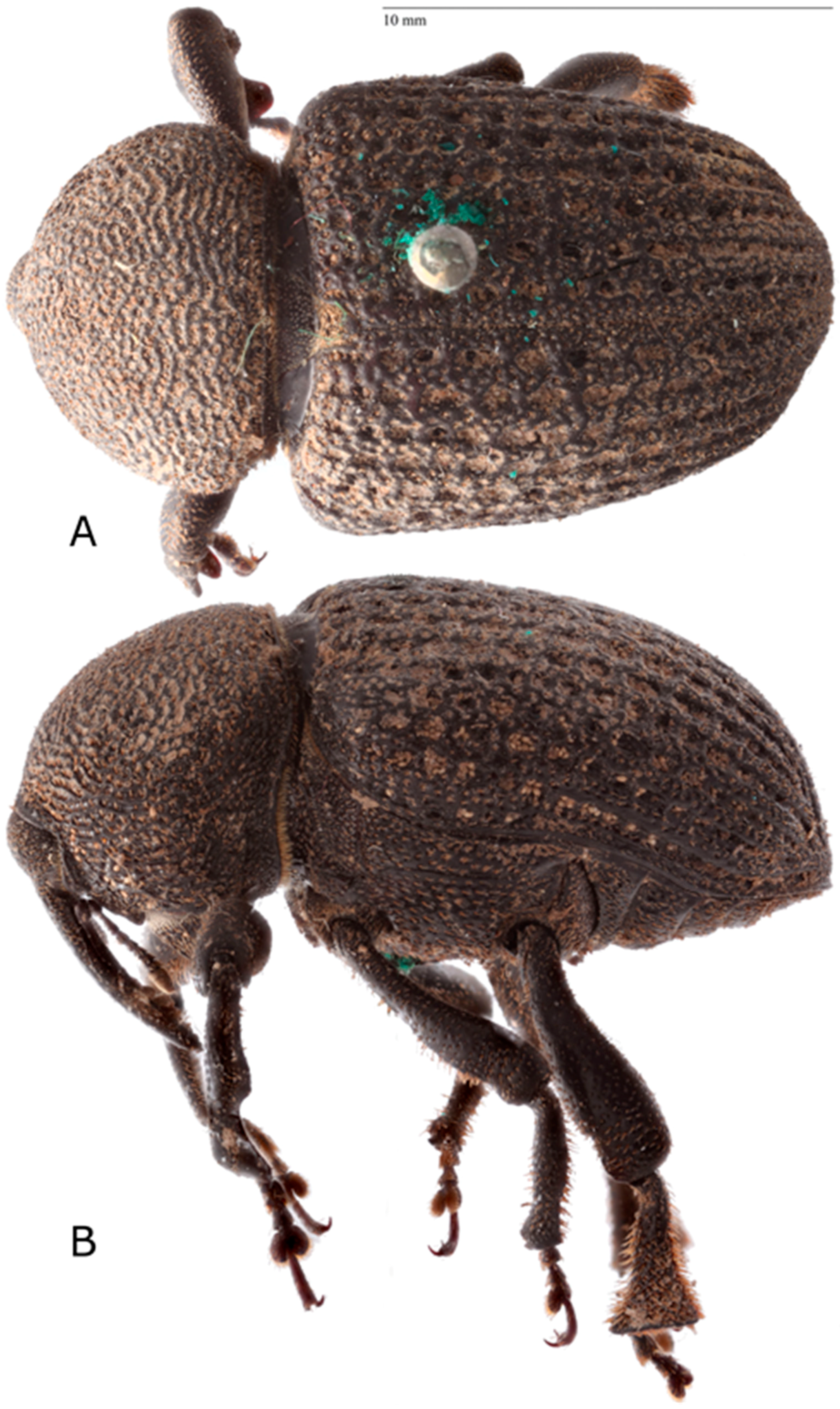
Sclerocardius africanus (Boheman) lectotype: (A) habitus, dorsal, (B) habitus, lateral. Photographed by Gunvi Lindberg (© 2018 Naturhistoriska riksmuseet). Original photo cropped, light levels and contrast adjusted. Made available by the Swedish Museum of Natural History under Creative Commons Attribution 4.0 International Public License, CC-BY 4.0.
Figure 1.
Sclerocardius africanus (Boheman) lectotype: (A) habitus, dorsal, (B) habitus, lateral. Photographed by Gunvi Lindberg (© 2018 Naturhistoriska riksmuseet). Original photo cropped, light levels and contrast adjusted. Made available by the Swedish Museum of Natural History under Creative Commons Attribution 4.0 International Public License, CC-BY 4.0.

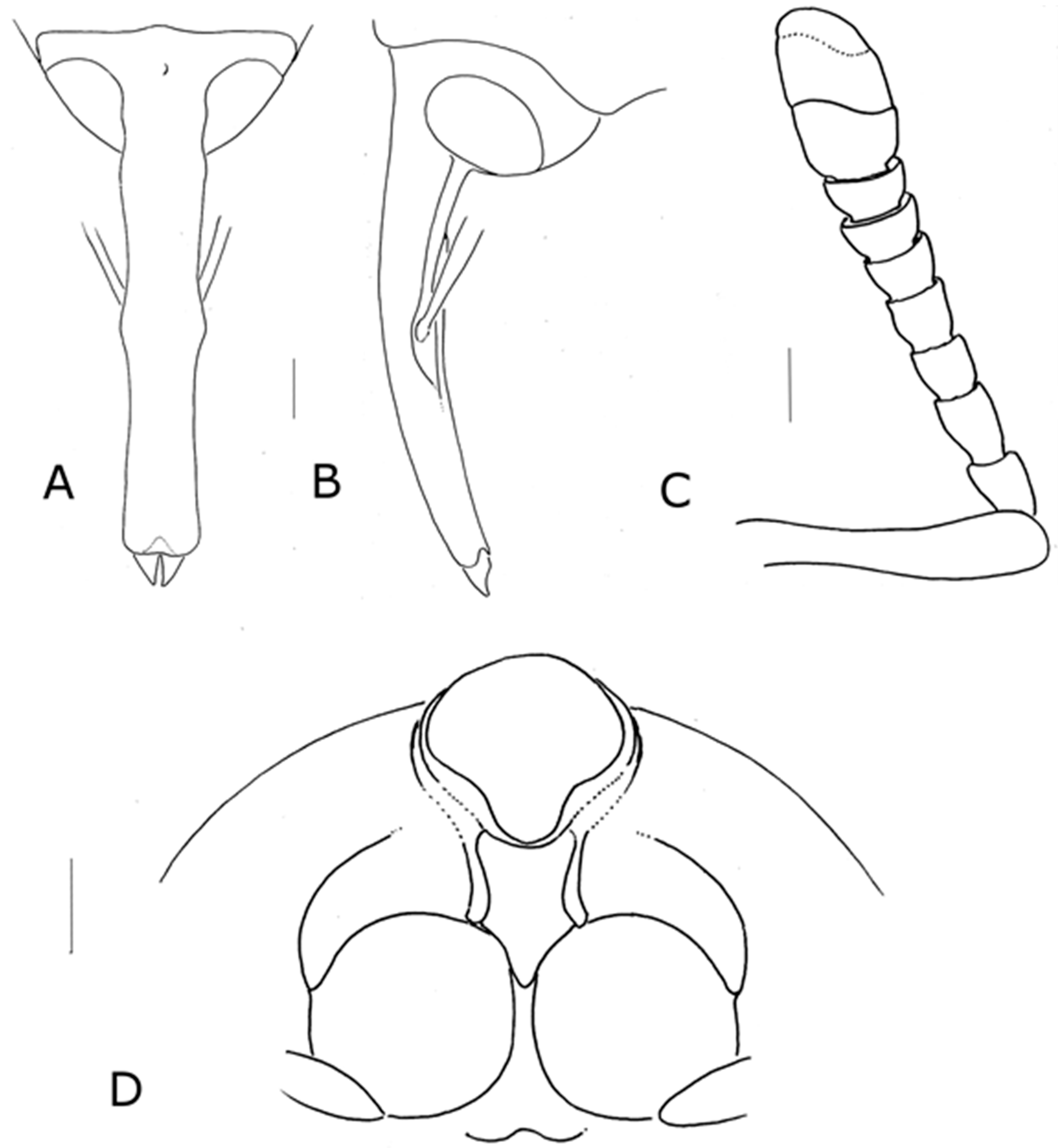
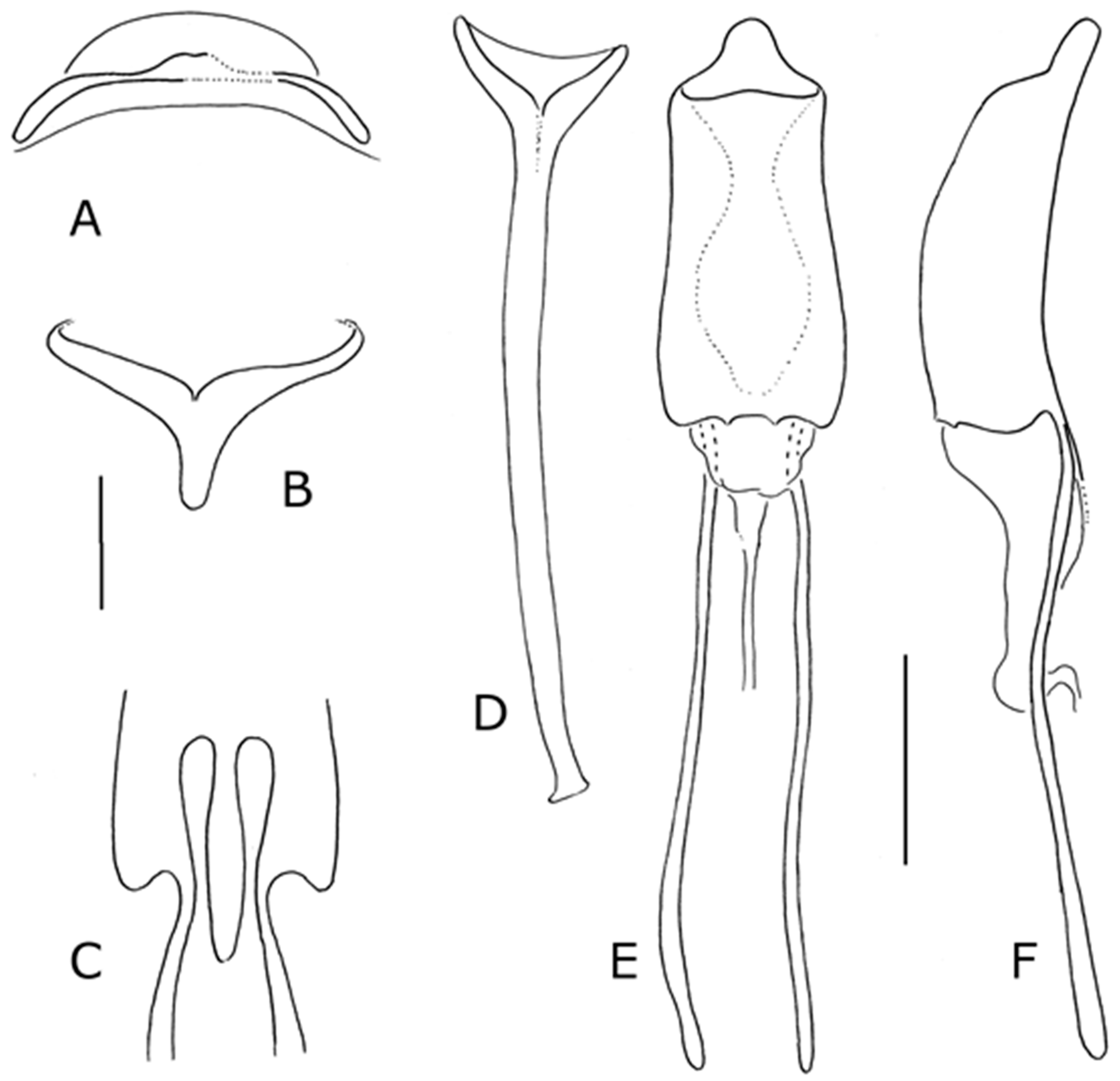
Figure 2.
Sclerocardius bohemani Schoenherr: (A) head dorsal (scale bar = 0.5 mm), (B) head lateral (scale bar = 0.5 mm), (C) antenna dorsal (scale bar = 0.5 mm), and (D) prothorax, ventral (scale bar = 1 mm).
Figure 2.
Sclerocardius bohemani Schoenherr: (A) head dorsal (scale bar = 0.5 mm), (B) head lateral (scale bar = 0.5 mm), (C) antenna dorsal (scale bar = 0.5 mm), and (D) prothorax, ventral (scale bar = 1 mm).

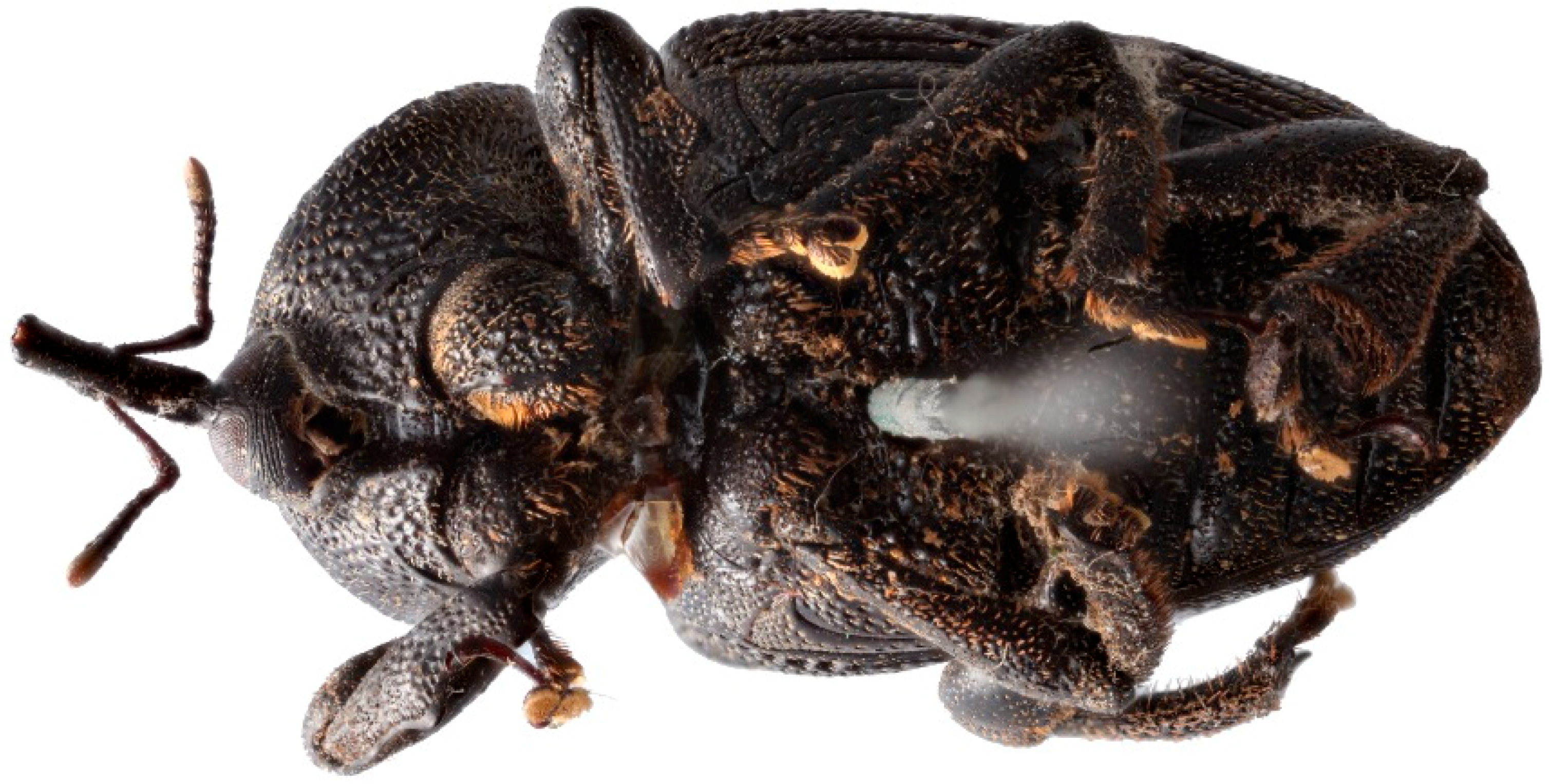
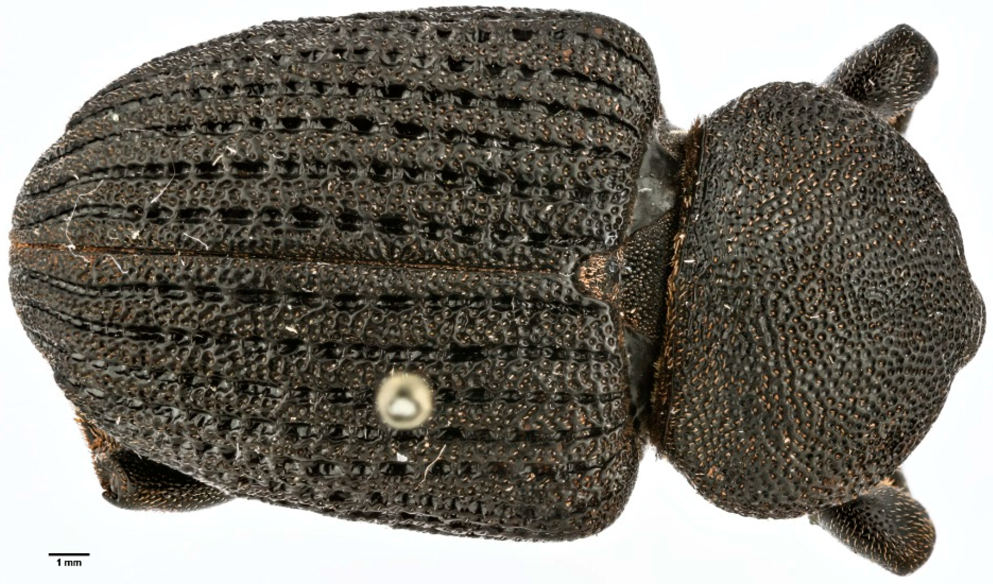
Figure 3.
Sclerocardius bohemani Schoenherr: head and prothorax, anterior (holotype of Charactocnemus hintzi Hartmann). Photograph by Marc Srour (Museum für Tierkunde).
Figure 3.
Sclerocardius bohemani Schoenherr: head and prothorax, anterior (holotype of Charactocnemus hintzi Hartmann). Photograph by Marc Srour (Museum für Tierkunde).

Figure 4.
Sclerocardius spp., elytra: (A) Sclerocardius africanus, and (B) Sclerocardius kuscheli.

Figure 5.
Sclerocardius africanus (Boheman): lectotype, ventral. Photographed by Gunvi Lindberg (© 2018 Naturhistoriska riksmuseet). Original photo cropped, light levels and contrast adjusted. Made available by the Swedish Museum of Natural History under Creative Commons Attribution 4.0 International Public License, CC-BY 4.0.

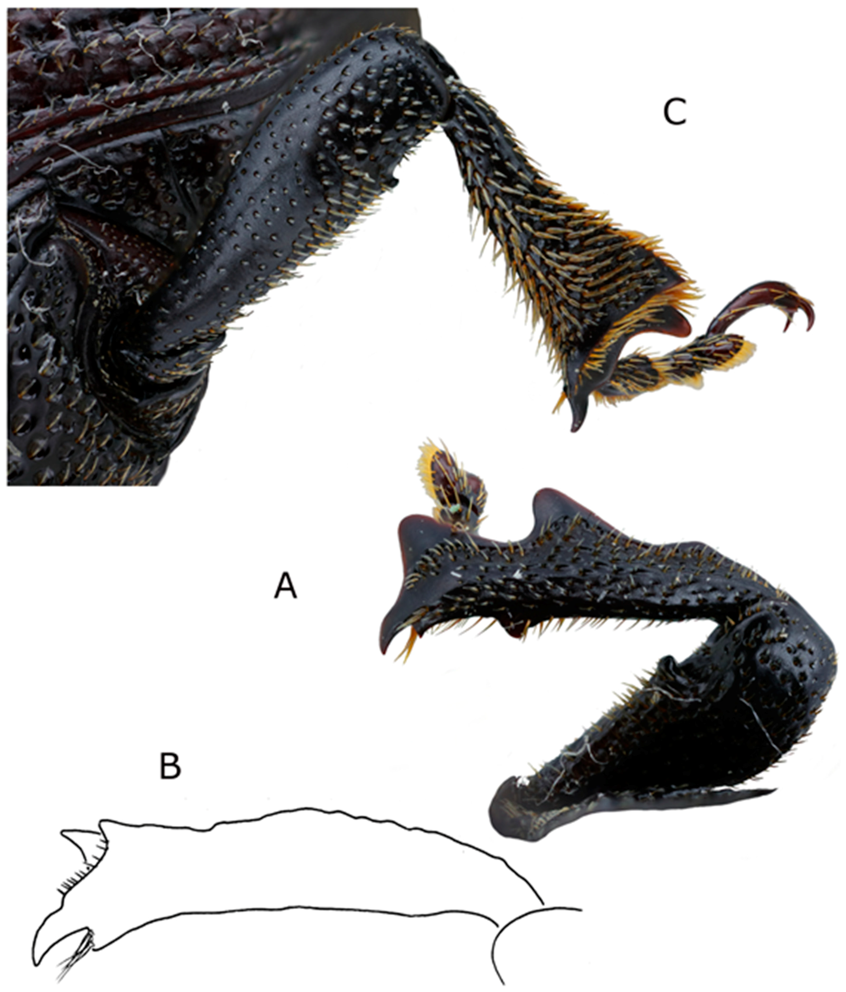
Figure 6.
Sclerocardius africanus (Boheman): (A) fore tibia, male right showing postero-ventral tooth, (B) mid tibia, right antero-dorsal, and (C) hind tibia left anterior.
Figure 6.
Sclerocardius africanus (Boheman): (A) fore tibia, male right showing postero-ventral tooth, (B) mid tibia, right antero-dorsal, and (C) hind tibia left anterior.

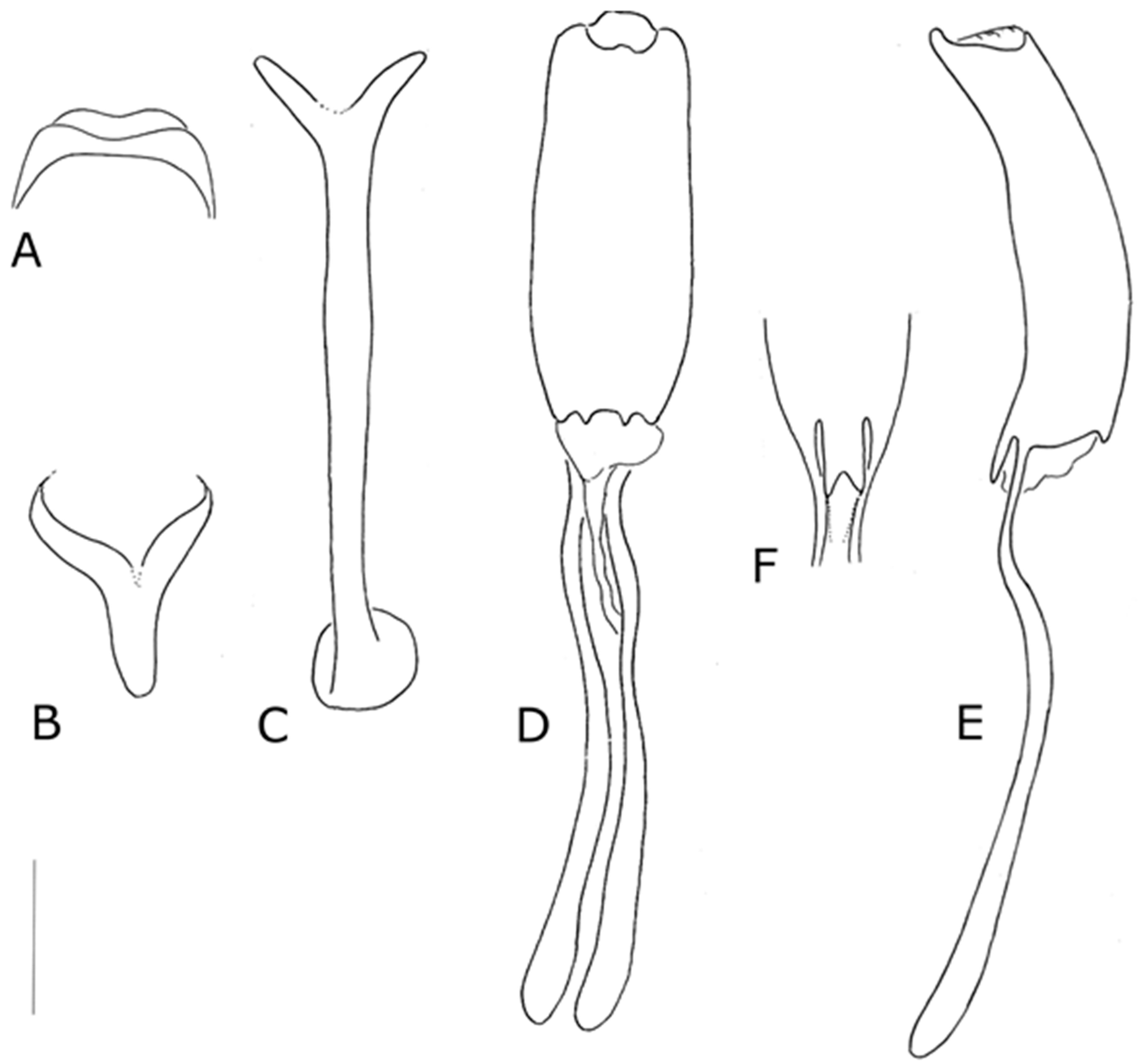
Figure 7.
Sclerocardius africanus (Boheman): (A) tergite VII male, and (B) tergite VII female. Scale bars = 1 mm.
Figure 7.
Sclerocardius africanus (Boheman): (A) tergite VII male, and (B) tergite VII female. Scale bars = 1 mm.

Figure 8.
Sclerocardius africanus (Boheman), male terminalia: (A) sternite VIII, ventral, (B) tegmen, dorsal, (C) spiculum gastrale, ventral, (D) penis, dorsal, (E) penis, lateral, and (F) penis, anterior ventral margin. Scale bar = 1 mm.
Figure 8.
Sclerocardius africanus (Boheman), male terminalia: (A) sternite VIII, ventral, (B) tegmen, dorsal, (C) spiculum gastrale, ventral, (D) penis, dorsal, (E) penis, lateral, and (F) penis, anterior ventral margin. Scale bar = 1 mm.

Figure 9.
Sclerocardius africanus (Boheman), female terminalia: (A) tergite VIII, dorsal, (B) tergite VII, lateral, (C) spiculum ventrale, ventral, (D) gonocoxites, ventral, and (E) genitalia, lateral. Scale bar = 1 mm.
Figure 9.
Sclerocardius africanus (Boheman), female terminalia: (A) tergite VIII, dorsal, (B) tergite VII, lateral, (C) spiculum ventrale, ventral, (D) gonocoxites, ventral, and (E) genitalia, lateral. Scale bar = 1 mm.

Figure 10.
Sclerocardous bohemani Schoenherr lectotype: (A) dorsal, and (B) lateral. Photographed by Gunvi Lindberg (© 2018 Naturhistoriska riksmuseet). Original photo cropped, light levels and contrast adjusted. Made available by the Swedish Museum of Natural History under Creative Commons Attribution 4.0 International Public License, CC-BY 4.0.

Figure 11.
Sclerocardius bohemani Schoenherr: holotype of Charactocnemus hintzi Hartmann, dorsal. Photograph by Marc Srour.
Figure 11.
Sclerocardius bohemani Schoenherr: holotype of Charactocnemus hintzi Hartmann, dorsal. Photograph by Marc Srour.

Figure 12.
Sclerocardius bohemani Schoenherr: (A) fore tibia, right (dotted line indicates smooth asetose areas), (B) Mid tibia, right anterior (setae omitted other than on premucro and apical comb), (C) hind tibia, right anterior, (D) tergite VII, male, and (E) tergite VII, female. Scale bars = 1 mm.

Figure 13.
Sclerocardius bohemani Schoenherr, terminalia: (A) male sternite VIII, (B) spiculum gastrale, ventral, (C) tegmen, dorsal, (D) penis, dorsal, (E) penis, lateral, (F) penis, anterior ventral margin, showing truncated form of projection, (G) penis, anterior ventral margin, showing complete form of projection, and (H) female spiculum, ventrale ventral. Scales bars: (A–E) 1 mm, and (F,G) 0.5 mm.

Figure 14.
Sclerocardius kuscheli sp.nov. habitus: (A) dorsal, and (B) lateral.

Figure 15.
Sclerocardius kuscheli sp.nov. tibiae: (A) fore tibia, right, (B) hind tibia, anterior right, (C) hind tibia, dorsal right, (D) fore tibia, antero-apical, to show premucro (arrowed), (E) mid tibia, anterior left, and (F) hind tibia, anterior right. Scale bar (line drawings only) 1 mm.
Figure 15.
Sclerocardius kuscheli sp.nov. tibiae: (A) fore tibia, right, (B) hind tibia, anterior right, (C) hind tibia, dorsal right, (D) fore tibia, antero-apical, to show premucro (arrowed), (E) mid tibia, anterior left, and (F) hind tibia, anterior right. Scale bar (line drawings only) 1 mm.

Figure 16.
Sclerocardius kuscheli sp.nov., tergite VII: (A) female, dorsal, (B) female showing wing-binding patch, and (C) male, dorsal (Scale bar = 0.5 mm).
Figure 16.
Sclerocardius kuscheli sp.nov., tergite VII: (A) female, dorsal, (B) female showing wing-binding patch, and (C) male, dorsal (Scale bar = 0.5 mm).

Figure 17.
Sclearocardius kuscheli sp.nov. male terminalia: (A) sternite VIII, ventral, (B) spiculum gastrale, ventral, (C) tegmen, dorsal, (D) penis, dorsal, and (E) penis, lateral. Scale bar = 0.5 mm.
Figure 17.
Sclearocardius kuscheli sp.nov. male terminalia: (A) sternite VIII, ventral, (B) spiculum gastrale, ventral, (C) tegmen, dorsal, (D) penis, dorsal, and (E) penis, lateral. Scale bar = 0.5 mm.

Figure 18.
Sclerocardius kuscheli sp.nov., female terminalia: (A) tergite VIII, dorsal, (B) tergite VIII, lateral, (C) spiculum ventrale, ventral, and (D) genitalia, ventral. Scale bar = 0.5 mm.
Figure 18.
Sclerocardius kuscheli sp.nov., female terminalia: (A) tergite VIII, dorsal, (B) tergite VIII, lateral, (C) spiculum ventrale, ventral, and (D) genitalia, ventral. Scale bar = 0.5 mm.

Figure 19.
Sclerocardius indicus Hartmann habitus, (A) dorsal, and (B) lateral.

Figure 20.
Sclerocardius indicus Hartmann, tibiae: (A) Fore tibia, right dorso-posterior (dotted line indicates smooth asetose areas), (B) fore tibia, anterior, showing postero-ventral teeth of male, (C) mid tibia, anterior left, and (D) hind tibia, anterior. Scale bar = 1 mm.
Figure 20.
Sclerocardius indicus Hartmann, tibiae: (A) Fore tibia, right dorso-posterior (dotted line indicates smooth asetose areas), (B) fore tibia, anterior, showing postero-ventral teeth of male, (C) mid tibia, anterior left, and (D) hind tibia, anterior. Scale bar = 1 mm.

Figure 21.
Sclerocardius indicus Hartmann, tergite VII, dorsal: (A) male, and (B) female. Scale bar = 1 mm.
Figure 21.
Sclerocardius indicus Hartmann, tergite VII, dorsal: (A) male, and (B) female. Scale bar = 1 mm.

Figure 22.
Sclerocardius indicus Hartmann, male terminalia: (A) sternite VIII, ventral (not fully pigmented in the figured specimen), (B) tegmen, dorsal, (C) penis, anterior ventral margin, (D) spiculum gastrale, ventral, (E) penis, dorsal, and (F) penis, lateral. Scale bars 1 mm ((A–C) at the same scale, (D–F) at the same scale).

Figure 23.
Sclerocardius indicus Hartmann, female terminalia: (A) tergite VIII, dorsal, (B) tergite VIII, lateral, (C) spiculum ventrale, ventral, (D) genitalia, lateral, and (E) gonocoxites, ventral. Scale bar for (A–D) = 1 mm, for E = 0.5 mm.
Figure 23.
Sclerocardius indicus Hartmann, female terminalia: (A) tergite VIII, dorsal, (B) tergite VIII, lateral, (C) spiculum ventrale, ventral, (D) genitalia, lateral, and (E) gonocoxites, ventral. Scale bar for (A–D) = 1 mm, for E = 0.5 mm.

© 2018 by the author. Licensee MDPI, Basel, Switzerland. This article is an open access article distributed under the terms and conditions of the Creative Commons Attribution (CC BY) license (http://creativecommons.org/licenses/by/4.0/).
Share and Cite
MDPI and ACS Style
Lyal, C.H.C. The Problematic Genus Sclerocardius (Coleoptera: Curculionidae: Molytinae: Ithyporini). Diversity 2018, 10, 74. https://doi.org/10.3390/d10030074
AMA Style
Lyal CHC. The Problematic Genus Sclerocardius (Coleoptera: Curculionidae: Molytinae: Ithyporini). Diversity. 2018; 10(3):74. https://doi.org/10.3390/d10030074
Chicago/Turabian StyleLyal, Christopher H. C. 2018. "The Problematic Genus Sclerocardius (Coleoptera: Curculionidae: Molytinae: Ithyporini)" Diversity 10, no. 3: 74. https://doi.org/10.3390/d10030074
Note that from the first issue of 2016, this journal uses article numbers instead of page numbers. See further details here.